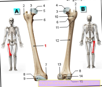
Illustration of the right thigh bone: from the front (A) and from the back (B)
Thighbones
Femur, os femoris
- Thigh shaft -
Corpus femoris - Great Rolling Hill -
Greater trochanter - Femoral neck -
Collum femoris - Femoral head (femoral head) -
Head femoris - Headband pit -
Fovea capitis femoris - Small rolling hill -
Lesser trochanter - Inner femoral gnar -
Medial epicondyle - Outer femoral gnar -
Lateral epicondyle - Articular surface for the kneecap -
Facies patellaris - Bone crest between
the rolling hills -
Crista intertrochanterica - Roughness for the approach of the
gluteus muscle -
Gluteal tuberosity - Rough line -
Linea aspera - Inner articular knot -
Medial condyle - Intergranular pit -
Intercondylar fossa - External articular knot -
Lateral condyle
You can find an overview of all Dr-Gumpert images at: medical illustrations
$config[ads_text1] not found
Tags:
Drug Psychiatry-Online Schnheitschirurgie Naturopathy Urology-Online