
Synonyms: Tendo calcaneus (lat.)
The structure known as the Achilles tendon is the insertion tendon of the three-headed muscle (Triceps surae muscle) of the lower leg. It is the thickest and strongest tendon in the human body.

The Achilles tendon is the thickest and strongest tendon in the human body. A tendon is the part of a muscle that connects the muscle to the bone and is made up of connective tissue. The Achilles tendon measures about 20 centimeters in length, is covered by a tendon sheath and consists of several tendon bundles, which in turn are composed of connective tissue fibers. The triceps surae muscle consists - as the name suggests - of 3 muscle heads. Two of them belong to the calf muscle (Gastrocnemius muscle), one of which belongs to the clod muscle (Soleus muscle).
$config[ads_text1] not found
All three muscle heads unite in their course to form an Achilles tendon, with which they are attached Calcaneus (Calcaneus) apply. The Achilles tendon sets on the entire width of the one located here Osseous prominence, the Calcaneal tuberosity, at. The Achilles tendon pulls over the upper part of this protruding bone and then attaches a little further down to the bone. So that the tendon does not lie directly on the bone in this area, is located here a bursa between the Achilles tendon and the bone (Bursa tendinis calcanei). A Bursa is a small liquid-filled sachetthat serves to the Reduce pressure and friction between tendons, muscles and bones.
At the base of the bone is the Achilles tendon widest, in the course upwards it tapers. The the narrowest point is about 4cm above the bone base, the so-called ´´Achilles tendon waist´´, then it runs in the three-headed calf muscle, becoming wider and wider. This is made up of two individual muscles: a two-headed calf muscle (gastrocnemius muscle), which arises on both sides of the thighbone (femur) in the hollow of the knee, and a single-headed clot muscle (soleus muscle). The clod muscle has its origin at the back of the Tibia (Tibia) as well as on Fibula head (Fibula).
The Achilles tendon transmits the force of this large three-headed calf muscle. This is especially the powerful flexion of the Foot towards the sole of the foot (Plantar flexion) and the Elevation of the inner edge of the foot with simultaneous lowering of the outer edge (Supination) made possible. The Achilles tendon runs at a certain distance from the lower leg bone, embedded between the superficial and the deep sheet of the so-called lower leg fascia, an enveloping layer of connective tissue. Also enclosed by these two fascia sheets is one Fat body (Corpus adiposum subachilleum), the Fills the space between the Achilles tendon and the lower leg bone. The skin above the Achilles tendon is relative thin and easy to move, hence the Achilles tendon itself easily palpable from the outside. The branches of the posterior tibial artery (Arteria tibialis posterior) and the Fibula artery (Fibular artery) supply the Achilles tendon with blood. The Innervation of the three-headed calf muscle and the Achilles tendon takes place via the Tibial nerve (Nervus tibialis), which comes from the Sciatic nerve (Sciatic nerve) arises.
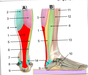
Here you get to: Illustration of the Achilles tendon
$config[ads_text3] not found
You can find an overview of all Dr-Gumpert images at: medical illustrations
Contracts the Triceps surae muscle, so this leads - via the Achilles tendon - to a Plantar flexion. That is the movement that one makes when one is on Tiptoe represents. Also at the Supination (turning the Footlike trying to see the bottom of the foot) the muscle with its Achilles tendon is involved.

Who am I?
My name is dr. Nicolas Gumpert. I am a specialist in orthopedics and the founder of .
Various television programs and print media report regularly about my work. On HR television you can see me live every 6 weeks on "Hallo Hessen".
But now enough is indicated ;-)
Athletes (joggers, soccer players, etc.) are particularly often affected by the disease of Achilles tendonitis. In many cases, the cause of the Achilles tendonitis cannot be identified at first. Therefore, the treatment requires a lot of experience. I focus on Achilles tendonitis.
The aim of every treatment is treatment without an operation with a complete restoration of performance.
Which therapy achieves the best results in the long term can only be determined after looking at all of the information (Examination, X-ray, ultrasound, MRI, etc.) be assessed.
$config[ads_text4] not foundYou can find me in:
Directly to the online appointment arrangement
Unfortunately, it is currently only possible to make an appointment with private health insurers. I hope for your understanding!
You can find more information about me at Dr. Nicolas Gumpert
The examination of the Achilles tendon reflex is part of the standard examination of the patient in neurology, among other things. Here the examiner stretches the muscle (and therefore also the tendon) of the patient usually suggests something by pushing / pulling the forefoot upwards a little and then striking the tendon with the reflex hammer.
If the reflex is intact, the foot moves towards the ground (Plantar flexion). The interconnection of the reflex runs via the tibial nerve and the spinal cord segments S1 and S2.
Achilles (Achilles) was a hero in Greek mythology who was mortal as the son of a divine mother and a human father. His mother Thetis wanted to at least make him invulnerable and for this purpose bathed him in the river Styx, which separated the underworld from the upper world. But the heel on which she held it when she dipped it did not come into contact with the water and thus became its only vulnerable place, the "Achilles' heel". Later it would be a poisonous arrow that hit and killed him right there.
It is particularly common in the Achilles tendon Tendon shorteningthat very painful could be. The best therapy ask for acute complaints stretching if possible several times a day and each for about 30 seconds. For example, you can use a simple Lunge forward, when the leg of the affected side is back, achieve a stretch of the Achilles tendon. To do this, straighten the upper body and bend the front leg, the rear leg is firmly on the floor and the heel is pressed down. Are suitable in everyday life Stairsto stretch the Achilles tendon by standing on the step with only your forefoot and slowly letting your affected heel hang down over the edge. If you experience pain while stretching, you should stop stretching immediately and seek advice from a physiotherapist. But also chronic inflammation the Achilles tendon, as is often the case with Athletes can occur through such Stretching exercises are treated and the with it the pain often associated with it subsides.
Under Taping one understands the application of a functional adhesive plaster tapethe treated Joints or Muscles does not completely immobilize, but only prevents unwanted or excessive movements.
Tape dressings can thus support the stability of the muscle and bone apparatus. Regarding the Achilles tendon can Taping help with pain, shortening and overloading, but plays a role especially in the prevention of Achilles tendon complaints.
So that a tape dressing can actually fulfill its function, it should be under professional guidance be created.
Strip-shaped tapes are affixed parallel to the Achilles tendon on the heel bone, the posterior fibula and under the foot to relieve the Achilles tendon and stabilize the ankle. The decision to use a tape bandage for the Achilles tendon should also always be made by a doctor or therapist.
A Kinesio tape bandage does not provide support for the Achilles tendon, it is not a substitute for a splint or achilles tendon bandage, it can only relieve discomfort and pain.
Acute injuries and swelling or external injuries speak against the use of a tape dressing and should be examined by a doctor or physical therapist. Also at Achilles tendonitis or partial tears of the Achilles tendon, taping is no longer sufficient.
Only in the final stage of recovery after therapy can a tape be used again as mechanical support for the Achilles tendon in order to stabilize the ankle.
You can find further information on this under our topic: Taping for Achilles tendonitis


.jpg "Hyperventilation (psychogenic)")


