Angiography is an imaging method used in medical diagnostics, in which blood vessels and connected vascular systems can be made visible. In most cases, with the exception of MRI, a contrast agent is injected into the vascular region to be examined. An image of the corresponding region is recorded using radiological recording methods, for example X-rays.

The contrast agent is distributed with the blood flow in the surrounding vessels and lights up in the X-ray image. This allows the vessel drawing to be precisely assessed in terms of position and course, as well as shape and pathological changes in the vessels.
Different types of angiography can be used depending on the vessel that needs to be examined. These differ in the type of contrast agent and the recording by an MRI, CT or Ultrasonic. The contrast agent is injected through a catheter after puncturing an upstream blood vessel. This puncture may be smaller Complications come.
$config[ads_text1] not found
In most cases, angiography provides precise information about the location and morphology of a blood vessel system. This allows the blood flow in the vessel and the supply of blood to a downstream organ to be assessed. For many important vascular diseases, both arterial and venous, angiography offers a precise diagnostic option.
Venous Thrombosis and Varicose veins can be visualized with venous angiography and their severity can be assessed. At Leg vein thrombosis the investigation is called Venography. This is where a blood clot blocks the flow in the vein. The angiography of the varicose veins is called Varicography. The superficial leg veins expand enormously due to a congestion of the blood.
In addition to vascular injuries, diseases in the arterial vascular system are primarily those arteriosclerosis, which is associated with vascular constrictions, and Aneurysms. An aneurysm is a bulging out of the arteries that can appear anywhere and, in the worst case, burst. Through angiography with Contrast media these vascular diseases can be represented in several images in such a way that both the morphology and the function of the vessel can be assessed. It also provides precise positional information, which is important for planning prior to operative vascular surgery interventions.
$config[ads_text2] not foundAngiography also gives you the opportunity to immediately after diagnostic imagingto perform an intervention. This can consist of widening a vessel, a Stent to set, treat an aneurysm, or remove blood clots.
DSA is an abbreviation for "Digital subtraction angiography". It is a variant of angiography in which the implementation remains the same, but the recording is digitally processed. The aim is to make interfering structures outside the vascular system invisible in the radiological image. This is possible by taking pictures before and after the injection of the Contrast agent. The computer digitally subtracts both images from each other so that only the contrast agent and thus the inside of the blood vessels can be seen.
By taking several images, even while the contrast agent is being introduced, a kind of film sequence results in which the agent can be seen spreading through the vessels. As a result, and by hiding interfering image aspects in the subtraction angiography, assessments of the form and function of the vessels can be made as precisely as possible. As a contrast medium at the DSA are predominantly radioactive iodine particles used, but newer methods can also be used with Saline solutions or work with CO2 as a contrast agent.
$config[ads_text3] not found
With angiography on the eye, the fine blood vessels of the Retina and the Choroid represent, which pull from the inside of the skull to the eyeball. Ophthalmologists make use of the angiographies on the eye if there is a strong suspicion of damage to the vessels.
$config[ads_text2] not foundTwo methods are available for angiography of the ocular vessels. They differ in the choice of contrast agent. It is the Fluorescence angiography and the Indocyanine green angiography. Both contrast media are available as harmless and harmless to classify. Before the examination is first carried out with special eye drop the pupil far put. The pupil is the only opening in the eyeball through which the vascular drawings of the retina can be seen. The respective contrast agent is then quickly applied via the arm vein. It only takes a few seconds for the contrast medium to reach the eye. The entire recording usually takes under 10 minutes.
The main causes that can damage the blood vessels in the eye are Diabetes, the Macular degeneration, the arteriosclerosis, Tumors or inflammation. With a very advanced diabetes mellitus it can lead to a "Diabetic retinopathy" come. In the case of macular degeneration, visual loss occurs especially in old age in the sharpest point of sight. Atherosclerosis, tumors and inflammation can attack the vascular structures in the eye and reduce or even stop normal blood flow. If the cells lying behind are not adequately supplied with blood, they will die Loss of vision would be the result.
Angiography of the eye offers a very precise diagnostic option for examining the blood supply to the retina. Unfortunately, it does not offer any possibility of direct intervention, for example removing a blood clot. It should be noted that the dilatation of the pupil means that there is a high sensitivity to light for a certain time after the operation.
$config[ads_text4] not found
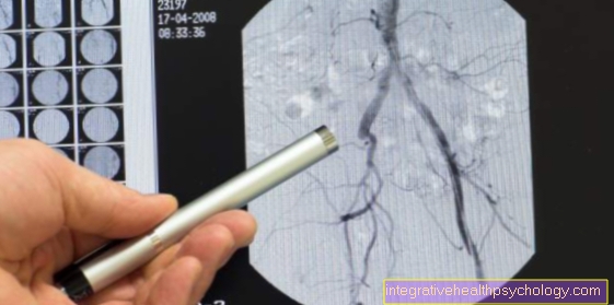
One of the most frequently used angiography examinations takes place on Heart instead of. The structures that contain blood are made visible here, i.e. the Coronary arteries and the Heart interiors even with right and left atrium and right and left ventricle. The consideration of the coronary arteries is also called "Coronary angiography" designated. Since the examination is carried out using a long catheter, it is also called a "cardiac catheter examination".
The catheter is a soft, flexible hosethat is slightly pre-curved at the tip to conform to the shape of the coronary arteries. The catheter is inserted from the outside into a more distant artery. An artery in the groin or elbow is used for this. In the opposite direction of the blood flow, the catheter is advanced through the vessel to the heart. It is made of a soft material so as not to damage the vessel walls.
With the help of a catheter one can also use pressure measurement techniques Contrast media inject into the vessels. This allows individual coronary vessels to be made visible and their function to be assessed independently of one another. The left ventricle of the heart, which pumps the oxygen-rich blood into the large body circulation, can also be enriched with contrast media. With simultaneous imaging, the Pumping power The left atrium and the left ventricle can be assessed, and cardiac catheterization through a vein is also possible. It is not used too often today. Here you can especially the right atrium and the right ventricle examine, but also the pulmonary arteries.
The angiographies are used on the heart for Assessing the size, Shape and pumping capacity both atria and both chambers. Other changes in the heart, such as tumors, Heart defect or calcifications can be detected. Coronary angiography, in particular, is crucial in diagnosing reduced blood flow to the heart, which is associated with health problems. Especially the CHD, Angina pectoris and Heart attacks represent consequences of arteriosclerotic changes.
The advantage of angiography on the heart is the direct intervention after the diagnosis. If the coronary arteries are narrowed, it is possible to use the catheter to widen the vessel, for example by inserting a Stents.
$config[ads_text1] not foundOne is usually fully conscious during the procedure. The most important thing after pulling the catheter out of the arterial vessel is that Stop bleeding with a tight pressure bandage. Complications from this procedure are rare.
Angiographies are generally a invasive diagnostic procedure. This means that the skin barrier is broken to get inside the body. The Complications are nevertheless manageable.
The most common undesirable complications are related to the puncture. Since that Contrast media must be injected into the blood vessels, a vessel is injured by a mostly very thin catheter. With arterial vessels this carries greater risks than with venous ones, since the Blood pressure is significantly higher in the artery. If the bleeding is not stopped sufficiently after the procedure, it will bleed into the tissue surrounding the puncture site. In rare cases, the vessel can also Aneurysms or Fistulas arise.
Depending on the location of the procedure, for example on the heart, a slight feeling of tension occur. You usually don't feel much during the exam itself. Theoretically, every part of the vessels and organs that the catheter passes can be injured by the plastic. Thanks to the particularly soft and flexible material of the catheter, these risks have largely been eliminated.
The contrast agent can be used in some people allergic reaction cause. Depending on the type of contrast agent, problems with the thyroid or the kidney occur.
The task of contrast media in angiography is through a radiological image different absorption behavior the X-rays to attract attention. In this way, the region through which the contrast medium flows can be clearly delimited from the rest of the soft tissue in the body.
Become particularly common iodinated contrast media used. Substances like Jor are also called x-ray positive designated. They absorb the radioactive rays to a high degree and thus form the contrast. Opposite them are various newer substances. These include Saline solutions or even gaseous carbon dioxide. They are called x-ray negative because they are extremely transparent to the rays. They are mainly used at Iodine intolerance.
For MRT angiography, so-called "Gadolinium chelates“Used.
Read more about this topic here: Contrast media
The angiography related to magnetic resonance imaging is also called Magnetic resonance angiography, short "MRA", designated. This creates a three-dimensional image in many layers and levels. There are several techniques whereby it is unlike other angiographic examinations it is not necessary to insert a catheter into the vessel. The great advantage of magnetic resonance angiography is that it can mostly without contrast media and thus manage without puncturing a vessel. The MRI, which measures the magnetization of all soft tissue, detects a high level of magnetization, especially when blood is freshly flowing in. Because the remaining tissues stand still and the blood flow only changes in the vessels, they can be displayed with a high level of signals.
$config[ads_text2] not found
In other procedures, a Contrast agent containing gadolinium be used. It is not necessary to use a long catheter. Even in very small quantities, it enormously enhances the appearance of the vessels. Another benefit of MRI is that too lack of radiation exposurethat at roentgen- or CT images always has to be considered.