Thoracic spine, thoracic spine, thoracic spine
The thoracic vertebrae are part of the human spine, start below the seventh cervical vertebra and end at the lumbar spine.
There are a total of twelve thoracic vertebrae in mammals, which are briefly numbered as Th1 to Th12. Th stands for the Latin term Pars thoracica "Chest part" of the thorax. Together with the ribs, they are involved in the structure of the thorax.
In general, the thoracic spine follows the construction principle of all vertebral bodies and serves as the base and origin of some muscles. The physiological shape of the thoracic spine is known as kyphosis, which is a backward convex curvature of the spine when viewed from the side of the body.
$config[ads_text1] not found
All vertebrae of the spine have the same construction principle. You own one Vertebral bodies (lat. Corpus vertebrae), as well as one Vertebral arch (lat. Arcus vertebrae).
Processes arise laterally and backwards from the vertebral body. The Transverse processes (Transverse process) go sideways and backwards Spinous processes (Spinous process). The spinous processes overlap like roof tiles and are easily palpable on the back.
The connection between the vertebral arch and the vertebral body forms the vertebral hole (Vertebral foramen). The successive vertebral holes together form the vertebral canal (lat. Vertebral canal) who the Spinal cord with its vessels, nerves and sheaths.
Between two vertebrae an intervertebral hole is formed, which allows the passage of the respective Spinal nerve allowed. Of the Pediculi arcus vertebrae, Vertebral arches, determine the bony boundary. The size of each Thoracic vertebrae increases from head to tail.
$config[ads_text2] not foundThe shape and the alignment of the joint surfaces also differ depending on the height of the thoracic spine. There are a total of six joint surfaces per thoracic vertebra. Two articular processes to the vertebra above, two to the vertebra below (lat. Processus articularis superior and inferior) and two articular surfaces to the Ribs (lat. Facies costalis).

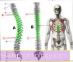
for a complete mapping of the thoracic spine

A - Fifth cervical vertebra (red)
B - sixth thoracic vertebra (green)
C - third lumbar vertebra (blue)
You can find an overview of all Dr-Gumpert images at: medical illustrations
$config[ads_text2] not found
$config[ads_text3] not foundThe individual sections of the spine differ in their Shape and size. Particularly noteworthy is the shape of the vertebral holes, which are in the Chest area are almost round and have the smallest diameter between Th 5 and 6 in contrast to the Cervical spine and Lumbar spine exhibit. Here the vortex holes are arranged in a triangle.
As already mentioned, the thoracic spine forms one convex curvature backwards that Kyphosis. The other areas of the spine form the opposite: the Lordosis.
The rib-vertebral joint is also a specialty Joint pits (Foveae costales superior et inferior) two thoracic vertebrae standing above or below each other take this Rib head on. Only the first, eleventh, and twelfth rib are excluded from this as they are only connected to one thoracic vertebra.
In addition, the laterally extending transverse process of the first to tenth thoracic vertebrae forms an articular surface that connects with the Rib humps (lat. Tubercula costae) is in communication. The eleventh and twelfth thoracic vertebrae do not form this articulated surface.
The last thoracic vertebra has like that Lumbar spine one Transverse process (= Transverse process) with a Mamillary process (in German: "Teat extension") and Accessor process (the additional appendix).
In addition, the rib and vertebral joint is through numerous ligaments stabilized.
The Forward and backward tilt is mainly about the ESPE carried out. The body can be bent about 45 ° forward and 26 ° backward. The thoracic vertebrae can be tilted sideways between 25-35 °. In addition, the Thoracic spine be rotated around their own axis. The circumference is about 33 °.
$config[ads_text4] not found
Generally, a anamnese, a conversation, led to an exact physical examination follows. The range of motion must be assessed here. There are two important tests for this. The Ott sign: From the seventh Cervical vertebrae a tape measure is placed in the vertical position of the patient and a line is marked 30 cm further down. Now the patient has to bend forward. The stretching of the vertebra is supposed to about 3-4 cm be. When flexing to the side, the finger-knee distance is measured.
Thoracic spine pain occur frequently and can have different types of pain. They are often described as dull between the shoulder blades or as belt-shaped pain in the Rib cage area.
The reasons for chest pain are many and varied; they can be the skeleton, muscles, or ligaments internal organs concern why a doctor should be consulted. Can be a cause of pain Herniated discs in the thoracic spine area be. However, they are very rare and can, with appropriate Pain therapy, as well as through anti-inflammatory and muscle relaxant drugs be treated.
Usually one also brings physiotherapy treatment an improvement in the symptoms. In rare cases this will be operational measures and only if the herniated disc presses on the spinal cord or nerves or there is a danger for one Paraplegia conceals.
Often enough for older people, especially women, due to osteoporosis, small trauma out to one Vertebral fracture trigger. Pain and immobility are common consequences.
As a therapeutic measure, the broken vertebra is straightened up again and filled with bone cement. This operation is called Balloon kyphoplasty designated. Sometimes this type of operation is not possible and the vertebrae must be stiffened (Spinal fusion). Adequate trauma must occur in young people to cause a fracture. Here, too, balloon kyphoplasty is primarily performed and only in the case of unstable fractures or significant kyphosis Stiffening operation necessary.
Approx. 15% of all Spinal fractures affect the thoracic spine. They mostly arise through High-speed trauma. The consequences are above all Compression fractures. Since the spinal canal generally has little reserve space at the level of the thoracic vertebrae, a narrowing of 20% by one is sufficient complete paralysis to evoke. The Spinal cord is affected in 2/3 of all injuries.
The extent of the injury is means various imaging (e.g. MRI of the thoracic spine) recorded and individually treated.
Conservative treatment is sufficient for stable fractures, but for unstable fractures, immediate surgery must be performed to restore the axes and stability, and to relieve the spinal cord.
In addition to the open surgical procedures are available today minimally invasive and thoracoscopic procedural techniques to disposal. The type of surgical treatment that is performed, however, depends on the type of fracture and the experience of the surgeon.
Another important clinical picture is the Scoliosis because it is particularly pronounced in the thoracic vertebra area. This is a extreme sideways inclination the spine, which can lead to some problems.

Who am I?
My name is dr. Nicolas Gumpert. I am a specialist in orthopedics and the founder of .
Various television programs and print media report regularly about my work. On HR television you can see me every 6 weeks live on "Hallo Hessen".
But now enough is indicated ;-)
The spine is difficult to treat. On the one hand it is exposed to high mechanical loads, on the other hand it has great mobility.
The treatment of the spine (e.g. herniated disc, facet syndrome, foramen stenosis, etc.) therefore requires a lot of experience.
I focus on a wide variety of diseases of the spine.
The aim of any treatment is treatment without surgery.
Which therapy achieves the best results in the long term can only be determined after looking at all of the information (Examination, X-ray, ultrasound, MRI, etc.) be assessed.
You can find me in:
Directly to the online appointment arrangement
Unfortunately, it is currently only possible to make an appointment with private health insurers. I hope for your understanding!
Further information about myself can be found at Dr. Nicolas Gumpert