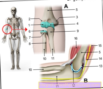
Medical: Articulatio cubiti
The elbow joint (Articulatio cubiti) connects the upper arm with the forearm. It consists of three partial joints, which are formed by three bones (upper arm, ulna and radius):
These partial joints are combined with a common joint capsule to form the elbow joint.
$config[ads_text1] not found
You can find an overview of all Dr-Gumpert images at: medical illustrations
$config[ads_text2] not found The elbow joint can be in two degrees of freedom be moved.
On the one hand, the forearm can be opened when the upper arm is not moving bend and stretch (Flexion / extension).
On the other hand, the elbow joint is functionally connected to the proximal radioulnar joint Turning movements involved in the hand (Pronation/Supination).
The main movements in the elbow joint are controlled by the Upper arm muscles executed.
The flexors (Flexors) are located on the front of the upper arm. These include:
The extensors in the elbow joint are on the back of the humerus. Also includes:
Individual forearm muscles are then also involved in pronation and supination.

Who am I?
My name is dr. Nicolas Gumpert. I am a specialist in orthopedics and the founder of .
Various television programs and print media report regularly about my work. On HR television you can see me live every 6 weeks on "Hallo Hessen".
$config[ads_text3] not found
As a former performance-oriented tennis player, I specialized early on in the conservative treatment of chronic tennis elbow.
In the past few years I have successfully treated several thousand tennis arms.
You can find me in:
Directly to the online appointment arrangement
Unfortunately, it is currently only possible to make an appointment with private health insurers. I hope for your understanding!
You can find more information about me at Dr. Nicolas Gumpert.
in the Humeroulnar joint (Articulatio humeroulnaris) form the "role" of the upper arm (Trochlea humeri) with a corresponding indentation on the ulna (Trochlear notch) a joint.
In close spatial connection to Trochlear notch, it says Olecranon, a protruding bone of the ulna that can be palpated as an "elbow".
The humeroulnar joint enables this Flexion and Extension (Flexion and extension) and is therefore a so-called Hinge joint.
The Humeroradial joint (Articulatio humeroradialis) is through the connection of the humerus head (Capitulum humeri) and the corresponding specialization (Fovea articularis radii) on the head of the spoke (radius head, Caput radii) educated.
This joint also has the two degrees of freedom the Flexion / extension to Flexion and extension of the forearm and the Supination / pronation to rotation of the hand.
$config[ads_text4] not foundStrictly speaking, this is a Ball joint. Ball joints always have three degrees of freedom (In addition to the degrees of freedom of the hinge joint, the abduction and adduction).
Since the humeroradial joint, however, by very strong band connections is secured, this last degree of freedom is omitted, so that it is anatomically a ball joint, which, however, only two degrees of freedom owns.
in the proximal radioulnar joint (Articulatio radioulnaris proximalis) are the edge of the radius head (Circumferentia articularis radii) and the corresponding notch on the inside of the Ulna (Incisura radialis ulnae) articulated together.
They form a so-called Wheel jointwhich one Rotation around the longitudinal axis of the bones makes possible. So that is this joint essential to the turning and turning movements of the hand involved.
The common large joint capsule closes all three partial joints and thus functionally combines them to form the elbow joint.
Attached is the joint capsule all three involved bone, i.e. on the upper arm, the spoke and the ulna.
In the area between the ring ligament (explanation follows) and the neck of the radial head, the joint capsule forms a bulge, the so-called protrusion Sacciform recess. This excess of capsular tissue serves as the Reserve fold and is used when the forearm is fully rotated in one direction.
The humeroulnar and humeroradial joints have strong band connections (Collateral ligaments) that lie laterally on the joint capsule.
These tapes (Ligamentum collateral ulnare and Radial collateral ligament) run in strong, fan-shaped stripsso that they support the joint laterally in every position:
The Ring band (Annular radial ligament) originates from the ulna, moves around the radius head and starts again from the ulna. This is how it secures the proximal radioulnar joint.
Bursa are liquid-filled, capsule-like delimited cavities, the outside the joint space lie and cushion heavy mechanical loads.
Bursae are either congenital or acquired (reactive bursae). Depending on the mechanical stress, bursae of different sizes develop in different places in each person.
Through this high individual variability you can no details to make the bursae of the elbow joint.
The largest bursa in the elbow joint is called Bursa subcutanea olecrani. It lies between the top of the ulna and the skin.
In the event of high mechanical stress or an open wound, a Bursitis come.
Read more on the subject: Tendinitis in the elbow.