
Most of the time, the symptoms of the disease tend to be unspecific Spondylolisthesis described. The clinical picture can usually not be diagnosed by the examination findings alone.
Only in the very advanced adolescent spondylolisthesis are gait changes (tightrope walk, sliding gait) or the jumping hill phenomenon. With the jumping hill phenomenon you can feel and see a jump-like deformation of the lower one Lumbar spine through vortex sliding.
Imaging tests help diagnose the disease and its extent.
$config[ads_text1] not found

Who am I?
My name is dr. Nicolas Gumpert. I am a specialist in orthopedics and the founder of .
Various television programs and print media report regularly about my work. On HR television you can see me every 6 weeks live on "Hallo Hessen".
But now enough is indicated ;-)
The spine is difficult to treat. On the one hand it is exposed to high mechanical loads, on the other hand it has great mobility.
The treatment of the spine (e.g. herniated disc, facet syndrome, foramen stenosis, etc.) therefore requires a lot of experience.
I focus on a wide variety of diseases of the spine.
The aim of any treatment is treatment without surgery.
Which therapy achieves the best results in the long term can only be determined after looking at all of the information (Examination, X-ray, ultrasound, MRI, etc.) be assessed.
$config[ads_text2] not foundYou can find me in:
Directly to the online appointment arrangement
Unfortunately, it is currently only possible to make an appointment with private health insurers. I hope for your understanding!
Further information about myself can be found at Dr. Nicolas Gumpert
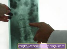
In principle, the X-ray of the spine as the basic imaging diagnosis of spondylolisthesis. The attending physician receives an insight into the posture of the spine via the X-ray images.
In addition, bony changes (calcium salt reduction, spinal curvature, a Vertebral fracture, Vertebral arthrosis (Facet syndrome), Vertebral body attachments, spondylolysis, spondylolisthesis) and disc subsidence can be recognized.
The spondylolisthesis can be seen on conventional radiographs on the side view.
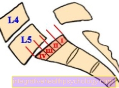
A severity classification of spondylolisthesis commonly used in clinics is that of Meyerding.
During diagnosis, the sliding process is divided into 4 degrees of severity, depending on the quarter in which the extension of the trailing edge of the sliding vertebra is on the sliding surface of the vertebra below
$config[ads_text3] not found
Some spinal instability cannot be seen on normal lateral images, but is only noticeable when the trunk is bent forward or backward. In these cases so-called help Functional recordings of the spine in forward and backward flexion of the trunk.
$config[ads_text2] not found
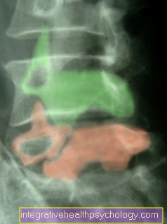
The spondylolysis defect can be identified in the Diagnosis best on oblique images of the lumbar spine as "Dog figure collar"Or in a computed tomography (CT) detect.
The sectional image diagnostics (CT and MRI, either with or without a contrast medium for the cervical / lumbar spine) enables the pain to be assigned to a specific nerve or a specific section of the spine.
With the help of a CT (C.omputertomography) examination of the spondylolisthesis, further questions regarding the bony structure can be answered (e.g. spondylolysis (vertebral sliding), spinal canal stenosis, vertebral body fracture).
However, this is even more valuable in spinal column diagnostics MRI of the cervical / lumbar spine (M.agnetrresonancetomography), which, in addition to the bony structures, clearly better than the CT, also the soft tissue structures (Band washers, Nerve roots, ligaments).
All of the above Diseases can be detected with the MRI and assigned to a specific section of the spine.
Read more about diagnostics under our topics:
$config[ads_text4] not found

The same X-ray image is shown twice in which the typical configuration of a spondylolysis (dog figure) can be seen.