Medical: substantia grisea spinalis
CNS, spinal cord, brain, nerve cells
English: spinal cord
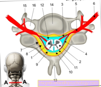
The shape of the gray spinal cord substance, which is butterfly-shaped in cross-section, can be divided into 10 layers (laminae spinales I-X) according to REXED.
Layers I-VI form the posterior horn / rear pillar (somatosensory = feeling), layers VIII and IX the front horn / front pillar (motor skills = muscles) and layers VII and X form a so-called "intermediate part" (pars intermedia) , in which various processing takes place.
The cells of the gray matter of the spinal cord can be divided into:
$config[ads_text1] not found
1st + 2nd spinal cord -
Medulla spinalis
You can find an overview of all Dr-Gumpert images at: medical illustrations
$config[ads_text2] not found
The Root cells are mostly motor nerve cells (nerve cells that control muscles) that leave the spinal cord via the anterior root. A distinction is made between different types of motor nerve cells:
The fibers of the skeletal and visceral muscles still contract in the anterior spinal root, but then separate.
The somatomotor root cells (= anterior horn cells, motor neurons) are the largest nerve cells in the spinal cord with a diameter of 40-80 m (that's 4-8 hundredths of a mm).
There are multipolar ganglion cells, which means that apart from an impulse-transmitting continuation (Axon) at least two "impulse-receiving" extensions (= Dendrites), but usually much more.
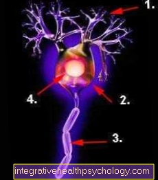
Many projections (axons) of other nerve cells end at them in the form of contact points (Synapses), the information from more distant parts of the body (periphery), from other spinal cord segments, from the Cerebral cortex, from the Cerebellum and from the Brain stem deliver. This information tells the motor neuron how to react in order to create a movement that is meaningful for the organism.
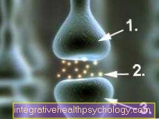
Figure nerve endings / synapse
The visceromotor root cells are smaller (15-50 m) and belong to the autonomous, so involuntary nervous system. They are also multipolar.
The cell bodies of the active in stress reactions Sympathetic nerve lie in the lateral horn of the thoracic and upper lumbar cords (C8-L2); their appendages (Axons) run briefly with those of the somatomotor anterior horn cells and then lead as so-called ramus communicans albus to the Sympathetic trunk (= Sympathetic trunk), next to the Spine runs. There they will be on a second Nerve cell switched.
The cell bodies of the active at rest Parasympathetic nervous system lie in the sacrum (= sacral) medulla (S2 to S4) between the anterior and posterior horns. Their appendages lead to Ganglia (= Accumulations of nerve cells) in the vicinity of their target organs, e.g. the intestines and other organs of the pelvis and lower abdomen, and are switched there.
$config[ads_text4] not found
The Internal cells receive nerve impulses from the sensitive nerve cells (Neurons), which are in the Spinal ganglia lie and their appendages (Axons) in the Back horn of the spinal cord. However, their appendages remain within the gray matter and convey the incoming information to different other nerve cells, depending on the cell type. The inner cells can be subdivided into
The Cells of the self-apparatus For the most part, as so-called inter-neurons (interneurons), nerve cells of the spinal cord connect with one another.
They are scattered in the gray matter of the spinal cord in different places. The
This own telephone ensures that on the one hand
If the skin experiences a sting, for example, the direct connections to the anterior horn cells result in defensive movements that still work when the spinal cord is separated from the brain by a cut.
Through cross-segment communication, all of the cells in the anterior horn that are required to move a muscle or muscle group can be reached, and the cross connections between the halves of the spinal cord also trigger a movement in the same direction on the other side: the reaction is bilateral.
For example, if we stumble with our left foot, reactions must still take place on both sides of the body in order to absorb the fall.
A simple one also works at this level Reflex path.
The lengths" Cord cells lie in the nuclei of the posterior horn of the spinal cord.
They belong to the afferents, that means to the ascending, supplying system: The cell bodies receive their information from the Spinal ganglion, which represents the first switching station (1st neuron) for sensitive information from inside the body and from the body surface, and thus form the second switching station (2nd neuron) on the way to the brain.
Their processes are long and form thick strands or tracts that rise to the brain. These run in the white matter on each side of the spinal cord anteriorly and laterally, in the so-called Front strands and Side strands.