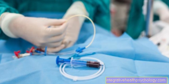
Coronary angiography
A cardiac catheter examination is understood to be a diagnostic or therapeutic measure to find and correct cardiovascular changes with the aid of a catheter inserted into the vascular system.

A Cardiac catheter is a very thin, inside hollow and several meters long instrument, in the central cavity of which there is a guide wire. This guide wire serves to pave the way for the actually rigid catheter (cardiac catheter).
The guide wire can be inserted and taken out variably. The tip of the catheter is slightly curved. If the guidewire is not inserted, the bend will remain at the tip. Is the Guide wire inserted, the bend at the tip is canceled. When the guide wire is pulled out, the catheter cavity offers the possibility of either injecting liquid in the form of contrast medium or further instruments right up to the catheter tip (Cardiac catheter) to advance.
$config[ads_text1] not found
Cardiac catheter examinations are now routine procedures for the reliable visualization of the heart vessels.
Thanks to modern technology, the procedure is relatively simple.
However, it is not free from complications. Mostly it concerns problems at the puncture site (Bruising etc.) which, however, do not require further treatment. Rarely can Complications at heart and other serious problems arise. However, this is more likely to be the case in emergencies, serious previous illnesses and an overall very poor general condition of the patient.
Contrast medium intolerance can occasionally occur. The cardiac catheter examination is therefore generally carried out on an outpatient basis on the awake patient with local anesthesia.
If no complications arise, the patient can leave the clinic on the same day. This applies to examinations without intervention. If there is secondary bleeding at the injection site, the patient remains usually overnight and can leave the clinic the next day without further complications.
More severe complications may require longer hospital stays. This rarely happens and depends on the type of problem and the general condition of the patient. Normally, patients should take it easy 3 to 4 days after the examination and stay in bed on the day of the examination.
Overall, cardiac catheterizations are performed on an outpatient basis if the course is uncomplicated.

Before one Cardiac catheterization (Cardiac catheter) some preliminary examinations must be carried out. They consist of one Resting ECG and one Exercise EKG, Blood count with Coagulation values Kidney and thyroid levelsto rule out that there is a contraindication for a contrast agent examination, and one X-ray the lungs.

goal of Cardiac catheterization is to the vascular system of the Heart to see and correct constrictions or occlusions. A cardiac catheter examination takes place in the so-called Cardiac catheterization laboratory instead, an operating theater-like room, which is kept particularly sterile on the one hand, and also has a bed next to it on the other X-ray machine Is provided. This X-ray device is attached in the form of an arch above the examination table and can be rotated around the patient.
To the Heart vessels To be able to make it visible, the catheter must be advanced into the heart. To do this, either puncture a peripheral vein (Right heart catheter) or one artery (Left heart catheter). The puncture of an artery is performed more often. Most often the inguinal artery is used as an access.
After finding the appropriate puncture site, a so-called lock used. These serve to keep the access open and at the same time to avoid bleeding caused by the high arterial pressure.
$config[ads_text2] not found
$config[ads_text3] not foundThe catheter (Cardiac catheter) slowly pushed forward through the vascular system. To pave the way, the guide wire is first pushed forward. It consists of a metal compound. During the advancement the examiner can through regular X-ray snapshots precisely determine the current position of the wire.
The destination of the cardiac catheter is the point of departure of the coronary arteries. These leave the Main artery just above the Aortic valve . As soon as the safe position of the wire is ensured by an X-ray, the blood vessels supplying the heart with oxygen-rich blood (Coronary arteries) shown. The catheter is pushed over the wire and a contrast medium is inserted through the actually hollow catheter Coronary arteries injected that quickly Heart muscle to distribute. The x-ray now shows in real time how the vascular system fills with contrast medium and how pervasive the blood vessel system is. Constrictions and occlusions are in the form of a Contrast medium recess clear.
During the examination, it is possible to document the examination and the results in the form of a video or photos.
Find yourself Constrictions of the coronary arteries, there is the option of expanding the vessel using a balloon introduced via the cardiac catheter and thus making it passable again. This method is also known as the PTCA (called percutaneous transluminal coronary angioplasty).
The balloon is passed limply over the cardiac catheter to the narrowed area and then unfolded. The pressure on the narrowed vessel causes it to widen.
There is also the option of a Stent insert into the narrowed or closed vessel. A stent is a tube made of a special material, resembling a wire mesh. A stent can also be introduced via the catheter probe (heart catheter) and pushed into the narrowed area. Similar to the balloon, it is pushed forward over the cardiac catheter in a folded state and unfolded after reaching the correct point. This keeps the vessel open.
Several stens can be inserted and several PTCAs performed in one catheter session. With completely closed vessels that lead to a Heart attack A stent is almost always used because it is more successful in keeping the vessel open.
$config[ads_text4] not found
At moderately to moderately narrowed vessels PTCA is often enough. In some cases it happens that a stent closes again after a while. In this case, the intervention must then repeated become. Newer materials are now available with one radioactive material coated. This is to ensure that deposits do not settle so quickly on the inner wall of the stent and can close it over time. Which material is used depends on the severity of the vascular disease, the condition of the patient and the examiner.
After Dilatation of the vessel and after the comprehensive radiological representation of the coronary system becomes the Cardiac catheter promoted back outside. The lock is pulled a few minutes later and given a pressure bandage. This may first 24 hours after the examination removed. In the meantime, however, one is working to shorten the duration accordingly. During this time, the patient should move and lie down as little as possible.
Before removing the bandage, the puncture site must be examined by a doctor. This listens to the areas above and next to the vessel with his stethoscope and checks whether Flow noise or a Hematoma is available. The pressure bandage can only be removed if the puncture site is not found. The reason for these precautionary measures is that the arterial system is under enormous pressure. Secondary bleeding are relatively common.
After a Stent implantation the patient should have a ASS-Clopidogrel Take a combination that is supposed to ensure that the blood remains thin and does not start to clot on the stent.

A Cardiac catheterization is always carried out when there is a suspicion of a acute coronary syndrome, Heart attack or Angina pectoris attack consists. Patients who report chest pain or pressure when exerted or at rest are potential candidates for cardiac catheterization. After a secured Heart attack (EKG changes, laboratory changes and patient's clinic) a cardiac catheterization is usually done to confirm and treat a heart attack. An examination will be carried out depending on the region and the availability of the nearest cardiac catheterization laboratory.
$config[ads_text1] not foundIf the next laboratory cannot be reached quickly enough, a drug lysis the blood will be thinned. However, there are numerous in large metropolitan areas in Germany Cardiac catheterization laboratories, and so this type of examination is the method of choice for heart attacks. Prolonged chest discomfort when moving (stable angina pectoris) or at rest (Unstable angina pectoris) can also be diagnosed and treated with a cardiac catheter examination (cardiac catheter).
A Cardiac catheterization (Cardiac catheter) should not be done if the patient has a strong one increased potassium levelsl or Digital mirror in blood has a infection or one sepsis is present, an uncontrolled high blood pressure or decreased blood pressure is present, a Contrast agent allergy exists, the patient is at one Renal failure suffers when he Problems with blood clotting has, or if a cardiac catheter examination has insufficient diagnostic or therapeutic value. Furthermore, no cardiac catheter examination should be carried out if a so-called Tachycardia (very fast pulse), one pronounced heart failure, one Inflammation of the heart valves or des Heart muscle or Heart sac exists or if the patient is in Pulmonary edema is located.
Please also read the article on the topic Contrast agent allergy.
The aim of cardiac catheter surgery is to Coronary arteries (Coronary arteries) or the heart itself using a contrast agent and X-ray technology to examine more closely. Cardiac catheter surgery works as follows.
First, the patient is prepared for the procedure in the cardiac catheterization laboratory.Since the doctor often accesses the Groin is used, it is shaved and thoroughly disinfected beforehand in order to minimize the risk of infection. Access via the Elbow or the Neck veins elected.
Then the bar locally anesthetized. This means that the patient remains during the entire cardiac catheter operation awareness and is approachable. The benefit here is that the patient can support the doctor during the procedure, for example with controlled breathing.
Once the local anesthetic has worked, the doctor may make a small incision in the groin and try to open the inguinal artery (Femoral artery) hold true. If this is successful, the bleeding is stopped and the catheter can be inserted into the artery. This is then pushed forward until it passes over the main artery (aorta) reaches the heart.
If the doctor now wants to examine the coronary arteries, he can pass the thin catheter through their outlets from the ascending main artery (Ascending aorta) lead and a Contrast media inject. This causes one in most patients Feeling hot in the chest, which, however, disappears after seconds.
Under constant fluoroscopy of the heart with an X-ray machine, the coronary arteries can now be made visible. The doctor can, for example, narrow them down (Stenoses) and if necessary with a balloon or Stent to treat.
$config[ads_text2] not found
Other important examination options in cardiac catheter surgery are checking the individual Heart valve function, the local measurement of Blood pressure and Oxygen content as well as a biopsy of the Heart muscle.
Read more about this topic here: biopsy
Once the cardiac catheter operation is complete, the wire is slowly pulled out of the patient's vessels and the access point is connected.
Complications occur with a cardiac catheterization extremely rare on (<1%).
If the incision in the groin is inaccurate, it can result from a nerve injury Sensory disturbances or paralysis of the leg come. Usually continue to occur harmless Arrhythmia (Extrasystoles) of the heart during the operation.
Depending on the age and diet of the patient, the catheter can also Cholesterol deposits in the aorta, which in serious cases leads to a stroke being able to lead. However, the incidence of such a complication is limited to less than one in every thousand cases.
Cardiac catheter surgery is a routine procedure that usually outpatient within someday can be carried out. The examination itself can vary depending on the condition of the patient's vessels half an hour and two last.
With positive results and subsequent treatment Cardiac catheter surgery can also be longer. Depending on the result of the procedure, the patient is often still allowed on the same day leave the hospital. After inserting a stent or a balloon, at least one additional day should be allowed for observation.
As with any procedure, in some cases (cardiac catheterization) complications can also arise from a cardiac catheter examination. Since the Cardiac catheter is advanced through the arterial vascular system into the heart, it is also in closer proximity Connection with the conduction system of the heartthat is responsible for every single heartbeat. If the Nervous system through the catheter tip it can be partly life-threatening arrhythmias which in most cases result in the examination being terminated.
A cardiac catheter is a foreign body in the arterial vascular system, on which, in extreme cases, the blood flowing past can clot. In this case there is a risk of Emoboliawhich can also have serious consequences (stroke, Heart attack, death).
Narrowed vascular sites come through in many cases atherosclerotic plaques accumulated over time due to a lack of exercise and an unhealthy diet. When expanding the vessel at this point it can lead to Tear open and to loosen plaques come, which can then be carried away with the bloodstream and clog a vital blood vessel elsewhere. An embolism could also develop here.
Furthermore, the cardiac catheter can injure the blood vessels of the heart and lead to bleeding. It happens often after a catheter examination Bleeding at the injection site. These should be avoided with a pressure bandage. Depending on the size of the hematoma, a surgical hematoma removal become necessary.
Also one Inflammation of the corresponding puncture site can be a result of the cardiac catheterization. The contrast agent can lead to intolerance reactions that make it necessary to stop the examination immediately.
The cardiac catheter is the gold standard in assessing the vascular structure of the heart.
This invasive examination alone enables a precise assessment of the vessels. In addition, the heart valves, congenital or acquired heart defects and other parameters of the heart can be assessed.
However, there are alternatives from which patients can also benefit in certain cases. Computed tomography of the heart (CT) and magnetic resonance imaging of the heart (MRI) are non-invasive options for examining the heart vessels. The advantage of both procedures is that they are not carried out invasively and are therefore without complications for the patient.
In addition, in contrast to cardiac catheters, the MRI does not provide any radiation exposure for the patient. However, the CT is associated with a certain radiation exposure.
Both procedures can indicate vasoconstriction. Experienced doctors decide individually whether a cardiac catheter examination is absolutely necessary for a patient. This depends on the type of previous illness, the suspected diagnosis and other factors. Whether an MRT or CT can alternatively achieve desirable results is also at the discretion of the doctor in the course of this assessment.
If the assessment of the functioning of the heart in general is to be checked, then the swallow echo can also be used.
The puncture site for the Introduction of the cardiac catheter usually takes place via a venous or arterial access to the groin, elbow or wrist.
Access to the wrist is transcarpal, which means via the wrist. There are then two possible arterial accesses, namely the Radial artery or the Ulnar artery.
The radial artery is located above the radius (thumb side), the ulnar artery is localized on the side of the ulna. The course of the investigation is similar to the usual procedure.