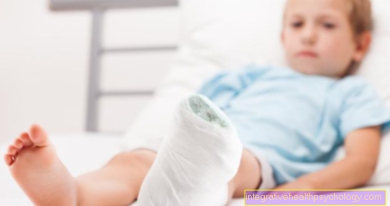
Medical: juvenile fracture
The human skeleton is particularly at risk of breaking bones (medical fractures) in childhood. This is due to the fact that the skeleton is still in the build-up phase at this time.
Thus the so-called growth plate (med .: epiphyseal plate), which is responsible for the longitudinal growth of the bone, is not yet closed. The outer and inner periosteum (endosteum and periosteum) are not yet fully developed either. These play a major role in the growth of bone thickness. The weak points of the child's skeleton are everywhere here.

$config[ads_text1] not found
If, in the event of a fracture, the bone parts are shifted sideways to one another, or rotated against one another, they are Misalignments spontaneously with growth in childhood corrected. Due to the not yet completed growth in length, the child's bone still has the potential to grow and thus compensate for the misalignment. In colloquial language one speaks of "fused" ("that fuses"). The extent of the potentially possible correction, however, varies depending on the age of the child, affected cooking and type of deformity.
After a fracture there may be something called a Spontaneous correction come.
However, if there is a bend in the axis in the event of a fracture, the correction is more difficult. The greatest potential for correction is in the forearm bones Cubit and spoke.
In contrast to lengthening - which can certainly be caused by fractures - which are not compensated for, shortening can sometimes correct itself spontaneously.
If a child has a broken bone, there is always one Risk of stunted growth. Especially with fractures of the bone shaft or bone parts near the Growth plate stimulation of the same leads to an extraordinary increase in length of the bone. In about 70% of children with an injury to the growth plate, the additional length growth is 1 cm.
$config[ads_text2] not found
If the epiphyseal plate (growth plate) is partially closed in the event of a fracture, this can lead to incorrect growth and shortening of the bones. The epiphyseal plate consists of several zones in which different growth processes take place.
The zone in the middle consists of cartilage and provides the cellular replenishment for the other cell zones (Reserve zone). The adjoining zone is the growth zone in which the cartilage cells form column-like. This is followed by the so-called Resorption zone. Here the cartilage cells enlarge due to water absorption. This process contributes significantly to the growth in length.
In this zone, the cartilage is solidified and forms factors that stimulate blood vessels to develop. Now comes the Ossification zone. The cartilage is replaced by bone. The increase in length is complete.
If the epiphyseal plate is closed, that too is Growth in length completed.
Particularly in childhood there are special fractures that are not found in adults due to the different bone structure. The bones in children are "softer".
Different types of fractions:
In the event of a compression fracture, compression occurs. That means the bone is being pressed together by force. The periosteum (periosteum) is preserved and does not tear when injured.
$config[ads_text3] not foundIn the case of a greenwood fracture, the bone breaks on the tension side and the compression side bends. The greenwood fracture gets its name because the child's bone has the property of breaking like a green twig. So it doesn't break through completely, but bursts apart without breaking.
$config[ads_text2] not found
Read more about the topic here: Greenwood fracture.
The Epiphyseal Injury is divided into Aitken and Salter. More on the classification of epiphyseal injuries can be found below in the text.

In the event of a Salter 1 injury, the growth plate loosens completely. With adequate therapy, the prognosis is good, but disturbances in bone growth are possible.
Solution epiphysis

A Salter 2 injury corresponds to an Aitken 1 injury.
Even with this type of injury, there is no injury to the growth plate, and disturbances in bone growth are also possible with this injury.
+ Break above the joint

A Salter 3 injury corresponds to an Aitken 2 injury.
The growth plate is involved in this type of injury. As part of the healing process, locally increased, but also decreased growth is possible.
+ Break below the joint

A Salter 4 injury corresponds to an Aitken 3 injury.
The growth plate is also involved in this type of injury. In the same way, with this bone fracture, a reinforced.
+ Break through the joint

In the case of a Salter 5 injury, which is not included in the Aitken classification, the growth structure is compressed without an actual bone fracture. Nevertheless, stunted growth can result.
Compression / bone bruise
You can find further information under our topic: Bone bruise
In the case of Aitken I or Salter I and II injuries, the epiphyseal plate remains intact. So the prognosis is good here.
In all other classification levels, however, the epiphysis is injured.
A childhood fracture has the same symptoms as an adult fracture.
Every fracture has different effects on the environment or the entire organism. Depending on the location, the effects can be more or less serious. If an adjacent organ is damaged by the fracture (for example, if the rib breaks, it could injure the lungs), the effects are more severe.
Pain is one of the Key symptoms of broken bones in children In addition to treating the fracture, it is therefore necessary to give the affected child a appropriate pain therapy to get rid of the pain. This is usually done by the Taking so-called NSAIDs reached.
Are the Bone fragments shifted against each other and it is followed by conservative therapy and a plaster cast, they must Fragments initially positioned become. Since this is a very painful procedure, one is often used Painkillers directly into the fracture line injected. In this way, the pain can be prevented from developing and the two fragments can be pulled into their natural position. When performing an operation, pain medication is also administered as part of anesthesia.
$config[ads_text1] not found
Depending on the fracture, it may be necessary to continue pain relief with NSAIDs for a few days after the operation or conservative care. As a rule, immobilization can relieve pain.
Fever can occur after a broken bone, especially in childhood. While fever in adulthood is almost exclusively associated with severe infections, it can Fever in children also occurs after a broken bone or other illness. This can have several causes. After a bone fracture, the body produces substances in the affected tissue that cause the destroy damaged body cells can. This process can lead to an increase in temperature in children. Also the Bruising which form as part of a broken bone are broken down and a reaction to this can result in a fever. Open fractures can cause a fever from infection and should be treated as soon as possible.
For Fever in children is infection in most cases responsible with a virus or bacteria. In contrast to adults, children are far more likely to be affected by infections. Sometimes the fever remains the only symptom of the infection. Not infrequently so is that coincidence of the fracture and fever and expression of a concomitant infection.

The most important measure for making a diagnosis is the X-ray. Sometimes x-rays of young children are difficult to assess because the bone substance has not yet consolidated. This fact may require an ultrasound of the affected area.
Of course, the appearance of the little patient and the associated medical history also play an important role. If the X-ray is not absolutely necessary, it should be avoided in children if possible.
Also read our topic: X-ray examination of the child
As a result, almost everyone Fracture pain, swelling and "bruises" called hematomas occur. Together with the limited functionality (e.g. resilience, mobility of adjacent joints), they are among the uncertain signs of fractures.
These include misalignment, abnormal mobility and so-called bone rubbing (a sound that occurs when parts of the fracture rub against each other).
The childlike skeleton is far from being mature. There is a high tendency to repair the bones. This tendency decreases with increasing age.
This tendency towards reparations justifies a conservative / non-surgical approach to child fractures in the case of uncomplicated fractures without significant malposition or damage to the Growth plate - they are usually put in plaster. The techniques depend entirely on the type of break.
In case of malalignment
Repositions - Return to normal position - must always be done under tension. The joining of the bones in one axis should be sought. They can also be compensated for at a later point in time with a wedge in the cast.
A plaster should the Wear time of 4 - 5 weeks do not exceed. If it is worn for more than 5 weeks, there is a risk that the muscles will recede too much.
The duration and type of plaster of paris always depends on the type of fracture and the healing process, so it can never be determined across the board.
In the Plastering of joints pay attention to the storage. Incorrect positioning can shorten the tendons and muscles and, after the fracture has healed successfully, the joint can be restricted in its function.
Surgical treatment is sometimes necessary in special cases.
For example:
The decision about conservative or surgical therapy must always be made taking into account all situational conditions.
Healing can be supported by homeopathy.
Read our topic: Broken bones and homeopathy.
Generally it is possible through the Taking homeopathic substances to support the healing of a broken bone in childhood. However, it must be noted that the Sole homeopathic treatment of the hernia cannot be recommended. Immobilization with a plaster cast or surgical therapy can ensure that no consequential damage occurs from the fracture and that the bone fragments heal well together. If in doubt, the treatment should be discussed with the treating orthopedic surgeon or trauma surgeon.
As Support bone healing should Calcium phosphoricum serve. This remedy is supposed to activate the metabolism of the bone substance and thus accelerate the healing of the bone. The recommended dose the remedy for broken bones in children is included D6.
The Duration until a child's broken bone heals completely depends on various factors. Both the Type of breakwhich bone is affected by the injury and which Type of therapy is decisive for the healing time.
In most cases, the conservative therapy with a plaster cast The therapy of choice is the plaster cast usually needs about Worn for 3-4 weeks become. Of the Complete healing of the bone is usually after 6 weeks to go out.
Will be a operative therapy The conservative method is usually the insertion of special wires, intramedullary nails or temporary external fixation. These procedures stabilize the bone so that A load on the injured bone is possible just a short time after the operation is. The introduced metals can be removed after about 3 months. The affected child is thus significantly less restricted, but the definitive healing of the bone still takes about 6 weeks.
Of the Forearm fracture in the child is one of the most common breaks in childhood. Both ulna and spoke can break here. Is the Broken bone in the middle, i.e. in the shaft area often becomes a Operation performed. In the distant part of the bone, on the other hand, conservative therapy with a plaster cast is often used. If the break occurs in the shaft area, the Duration up to 15 weeks for healing. In the wrist area, it rarely takes more than 5-6 weeks for the bone to heal completely. It is important here whether the growth plate is affected by the break. If this structure is involved, it depends on the type of fracture and the position of the bones whether treatment with a plaster cast is sufficient or whether surgical therapy is the method of choice.
Upper or lower leg fractures usually only occur after an enormous amount of force. This can be done, for example, by a Fall from a great height be the case. If the plant has a Plaster cast is necessary, this is for 3-5 weeks carried. Surgical treatment is also often performed in this area. One is not uncommon Relief of the affected leg with crutches or a wheelchair for several weeks necessary. Depending on the location of the fracture, it takes at least 6 weeks for the bone to heal. The metallic objects in surgical therapy are usually removed after about 3 months.
The duration up to Greenwood fracture healing depends on the type and location of the break. In order to improve the healing process, the Fracture affected bone completely broken under anesthesia become. Sometimes the break can be easy supplied with a cast be what about 3-5 weeks should be worn. However, it may also be necessary to fix the bones in place with wires. In this case, healing can take longer, but the bone is often load-stable immediately after the operation.
In children, the fractures heal more quickly than in adults. The risk of joint stiffening is also significantly lower. Any malpositions that may have occurred during the healing process can be compensated for by the increase in length (correction potential).
A special follow-up treatment (in general) is not necessary. The follow-up treatment is always based on the circumstances of the individual bone fracture.
However, attention should be paid to the early removal of any foreign material (wires, flaps, screws, etc.) that may have been introduced during an operation.
In order to be able to rule out growth disturbances with certainty, all fractures of the growth plate, all fractures of joints and legs must be checked. This control should extend over two years, but at least until the growth is complete.
The most common fracture of the arm in childhood is the so-called distal radius fracture (wrist fracture), so the break of the spoke right above that wrist.
Shaft fractures are 50 times more common than fractures of the epiphysis (growth plate).
Also Injuries from the elbow are very common. However, these are mostly Elbow dislocations, in particular to dislocations of the head of the spoke (med. radius head = Chassaignac dislocation). Dislocations are dislocations. The special radial head dislocation, in most cases, is not a complete dislocation that is medically considered to be Subluxation referred to as.
The medical name of this condition is: Chassaignac dislocation.
Children suffer from fractures of the bone shaft more often than the joints. Joint fractures are much more difficult to treat. However, they occur on the arms, especially the forearm, about twice as often as on the legs. Lower tibia fractures account for around 7 percent of fractures in childhood and adolescence.
You can find more information about a child's forearm fracture under our topic: childish forearm fracture.
Breaks in childhood are common. They are special because the little patients are still in the growth phase and so are their bones. As long as they grow, children have a so-called growth plate in their bones.
This can be used to classify fractures (Aitken and Salter). The severity of the break and the consequences can also be recorded in this way. The symptoms are the same as in adults:
In order to confirm the diagnosis, an x-ray usually has to be taken. In children, therapy can be conservative, i.e. with plaster of paris. Operations are only necessary in certain cases.
A specific follow-up treatment is not necessary. However, material introduced during the operation should be removed early. Children who have suffered high-risk fractures (fractures of the growth plate, joints or legs) should be examined regularly for growth disorders.