Synonym: Ventriculus sinister, left ventricle
The left ventricle is part of the "great" or body circulation, the left atrium (Atrium sinistrum) downstream and pumps the oxygen-rich blood freshly coming from the lungs into the aorta and thus into the body's circulation, where it supplies all important structures with oxygen.

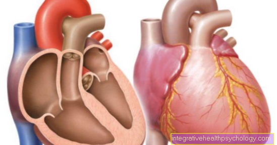
The heart lies rotated around its longitudinal axis in the left chest so that the right half of the heart more the anterior chest wall (ventral) is applied while the left half of the heart rather after back (dorsal) shows.
The left ventricle (left ventricle) is divided into an inflow and an outflow path. It is separated from the atrium by the bicuspid valve or mitral valve. This is through Tendon threads (Chordae tendineae) with the Papillary muscles connected that arise on the ventricular wall and ensure, that the Shut up at their end before and during the tension phase (Systole) the left ventricle does not strike back too hard into the left atrium.
The blood reaches the outflow path during systole after passing through the Aortic valve in the body circulation.
The ventricles (Ventricle) differ from each other due to their function:
$config[ads_text1] not found
The left and right ventricles are Chamber septum (Interventricular septum) separated from each other, the septum has a thickness of 5-10 mm.

You can find an overview of all Dr-Gumpert images at: medical illustrations
$config[ads_text2] not found
The heart will functionally divided into a left and a right heart. The right heart is part of the "great" circulation (body circulation), over four Pulmonary veins (Pulmonary veins) the blood reaches the left atrium and from there via the bicuspid valve (also: Mitral valve) into the left ventricle. After contraction of the left ventricle and opening of the aortic valve, the blood enters the Main artery (aorta), through which the blood flows through the body via various vessels and supplies it with oxygen (and other nutrients).
The heart action is roughly in two sections subdivided that diastole and the Systole. in the left heart this cycle takes place as follows:
This Heart action consisting of systole and diastole runs synchronously with it and on the same principle in the right ventricle from which the blood first enters the Pulmonary circulation is pumped. After having it there oxygen Once saturated, it enters the left atrium and the cycle of diastole and systole repeats again.
$config[ads_text2] not found
$config[ads_text3] not foundRead more on the topic: Task of the heart
The Wall layers are The same in all four interior heart rooms built up:
The heart gets over that Coronary arteries (Coronary vessels, vasa coronaria) supplied with blood. These are through the two main vessels, the left and right Coronary artery (Left and right coronary arteries) and their numerous branches.
This arise from the aorta, right after it leaves the heart. The left ventricle is mainly supplied by branches of the left coronary artery, but the right coronary artery also provides a small part of the supply.
A Insufficiency of the heart means one Muscle weakness, due to the Pumping power of the heart Not more sufficient, to provide the body with sufficient oxygen.
Thereby jams more and more Blood in front of the affected part of the heart.
At a Left heart failure this backwater mainly takes the form of a Pulmonary edema, i.e. a noticeable accumulation of water in the lungs. Typically this causes pulmonary edema Difficulty breathing (Dyspnea). Another symptom of heart failure is - among others - a Decline in physical performance. causes can be for example:
$config[ads_text4] not found
Ventricular septal defects are relatively common congenital malformationsthat have an opening in the ventricular septum. So there is one Short circuit (Shunt) between the left and right ventricle. This means that already oxygen-saturated blood from the left ventricle is not only ejected into the aorta, but also pumped back into the right ventricle (Left-right shunt). This leads to a increased stress on the left ventriclebecause it now has to do more work to pump the required volume into the body's circulation. This results in a Cardiac hypertrophy (Increase in thickness of the heart wall). To prevent this from happening, larger ventricular septal defects are made surgically corrected.