
Tonometry
English: intraocular pressure measurement
An intraocular pressure measurement is understood to mean different mechanisms for measuring and determining the pressure that is present in the anterior segment of the eye.
The intraocular pressure measurement, also known as tonometry, is a standard method for examining and detecting an intraocular pressure that may be too high glaucoma (glaucoma).
General information on the topic can be found here: Intraocular pressure
A slight pressure on the eyeball enables a first rough assessment of the pressure prevailing in the eyeball. Very strong deviations from normal pressure inside the eye can thus well recognized become.
$config[ads_text1] not found
At slight deviations or only moderately increased pressure in the eye gives this procedure alone, no information about the extent or severity of the disease.
Thus, for a precise estimation of the intraocular pressure, the measurement by means of Tonometer in the foreground.
The assessment of the intraocular pressure alone is not decisive and decisive for the development later Consequential damage, or the glaucoma to be mentioned primarily in this context.
However, too much pressure in the eye increases the risk of one later illness, With Loss of optic nerves and to get fibers, which, depending on their characteristics, can lead to more or less severe visual impairments for the person concerned.
The most important application of this examination is therefore the diagnosis of glaucoma. In addition, the further follow-up control in the event of increased values.
That means that you then get by one at regular intervals half year should have the intraocular pressure measured.
$config[ads_text2] not foundIf there is a family cluster, one Glaucoma As a precaution, regular examinations should continue to be carried out every year.
The examination can be carried out at Ophthalmologist.
Regardless of any illness or complaint, it is recommended to measure intraocular pressure from 40th year of life, in patients one glasses should get to perform.
Palpation:
Before there were corresponding instruments and apparatus for measuring intraocular pressure, intraocular pressure was determined using this method. The intraocular pressure measurement can also be carried out by any non-ophthalmologist today in order to obtain an overview of the pressure conditions inside the eye. With this method, the practitioner sits across from his patient.
The patient is asked to close their eyes, and the examiner gently and gently presses one of the eyeball with both index fingers while the rest of the fingers are supported on the patient's forehead. Depending on how far the surface of the eyeball can be pressed in, a rough estimate of the pressure conditions can be made. The intraocular pressure measurement must be carried out extremely carefully, but an exact pressure measurement is not possible with it. This examination method is particularly useful to diagnose a glaucoma attack in which the eyeball is not indentable and as hard as a board. The comparison between the sides is particularly important. A pressure difference between the left and right eye can indicate glaucoma.
$config[ads_text3] not found
Applanation tonometry:
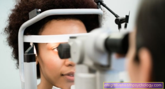
Applanation tonometry is performed on a measuring device called a tonometer. While sitting, the patient rests his chin on a mat and his forehead is pressed against a strap. The one opposite Ophthalmologist drives close to that with a small cylinder eye and very carefully places this cylinder on the patient's wide-open eye.
The applanation tonometry of the intraocular pressure measurement measures the force that is necessary to cover an area of 3mm Press in the diameter so that it is flattened. Once this has happened, the pressure applied corresponds to the intraocular pressure. The ophthalmologist sees two circles on his side of the device, which must be moved towards each other by turning a button (on the side of the tonometer) until they are on top of each other.
The intraocular pressure is then read off on a scale. Since the eye is sensitive to pain and irritation, it is necessary to numb the surface of the eye. A fluorescent liquid is also placed in the eye. The intraocular pressure varies in healthy people and also depends on various factors, such as the thickness of the cornea. The thicker the patient's cornea, the more pressure has to be applied to dent the surface, which is a formal one Increase in intraocular pressure that does not exist. For this reason, it is always necessary to determine the corneal thickness of the patient in the case of questionable high values.
Patients who are lying down can be examined using so-called hand applanation tonometers. Such mobile devices are also used for so-called day-night measurements, in which intraocular pressures also have to be measured at night.
Non-contact tonometry:
With this measuring method the Intraocular pressure measurement the device does not touch the cornea during the measurement. Instead of the cylinder, the cornea is flattened with a short, powerful blast of air. This creates a visible reflex that can be evaluated by the device and shows a corresponding intraocular pressure. Since there is no direct contact with the cornea, surface anesthesia of the cornea is not necessary. Possible risks of injuries to the cornea or infections are also minimized. The results of this intraocular pressure measurement are not as accurate as with theApplanation tonometry. For the patient, the non-contact tonometry is also the more unpleasant examination. The air blast measurement also only works with intact corneal surfaces. For scarred or injured corneas (Astigmatism and Corneal ulcer) incorrect values are displayed.
Impression tonometry
This is an older method of measuring intraocular pressure, in which a pen is placed on the cornea and then it is measured how far this pen dips into the corneal surface with its weight. The corresponding intraocular pressure is then determined from this. In this procedure, too, the cornea is anesthetized before the examination eye drop to treat. Today applanation tonometry and non-contact tonometry have largely replaced this method. This form of intraocular pressure measurement is still used in patients who have a scarred cornea and the first two measurement methods mentioned do not allow reliable values.
Overall, it has to be said that impression tonometry does not give precise values for intraocular pressure.
$config[ads_text4] not found
In addition to non-contact tonometry, the other intraocular pressure measurement methods still have some risks that should be observed. First of all, the patient may have an allergic reaction to the anesthetic drops previously placed in the eye. Burning eyes after the drop is normal and will go away after a few minutes.
An allergic reaction can, however, include systemic reactions, such as shortness of breath or anaphylactic shock. Furthermore, injuries to the cornea and the surface of the eye can also be caused with all measuring methods of intraocular pressure measurement in which direct contact with the cornea is established. Scratches and tears of the cornea from excessive pressure should be mentioned. In extreme cases, a corneal transplant may be necessary. Furthermore, when measuring intraocular pressure there is a risk of germs being transmitted, which is a Keratoconjunctivitis epidemica can trigger and require antibiotic treatment.

The most important indication for intraocular pressure measurement is the diagnosis and follow-up of a Green stars (glaucoma). The examination should be carried out from the age of 50 in order to find out new diseases. Depending on the result, the examination must be repeated at regular intervals. In the case of increased pressure values, the examination should take place every six months. If glaucoma has already occurred in the patient's family, an examination once a year is recommended.
The intraocular pressure measurement is a preventive examination and is usually not paid for by the health insurance company. It therefore falls into the category of so-called individual health benefits (IGeL), which everyone has to pay for themselves.
The costs amount to EUR 20, which the patient has to pay himself if there is no known glaucoma (preventive care). For all suspected glaucoma patients, the examination is carried out as a follow-up and is therefore covered by the health insurance company.
The Standard values the intraocular pressure are usually in the range of approximately 10 to 22 mmHg. Of the Average is approximately in the range of 15 mmHg. The amount of the value is depending on the time of day and is subject to fluctuations. The intraocular pressure is particularly high in the morning hours or after getting up the highest.
$config[ads_text1] not foundDaily pressure fluctuations of up to 4 mmHg are to be regarded and owned as normal no disease value. With values around 22 to 26 mmHg there is a suspicion of one glaucoma, so that in case of doubt, further intraocular pressure measurements must be carried out.
All measurement the values of over 26 mmHg are always to be assessed as pathological with regard to an existing glaucoma. This requires clarification of the cause and treatment and lowering of the pressure to avoid or minimize consequential damage.

Of the Intraocular pressure is built up in the anterior chamber, which extends between the cornea and lens in the anterior segment of the eye. The pressure is created by a balance between the production and outflow of aqueous humor and is maintained in a healthy patient. The aqueous humor is formed by the ciliary epithelium of the eye, then flows through the anterior eye area and finally reaches the venous blood system via Schlemm's canal. The intraocular pressure built up is necessary to maintain the shape of the eye and to ensure the refraction of light, among other things. The intraocular pressure increases when the outlets to the blood system are blocked. The risk of increased intraocular pressure lies in damage to the Optic nerves, on the fundus of the eye, which can only tolerate a certain pressure area without damage.
The normal eye pressure in humans is between 10 and 20 mmHg. There is a wide range of norms that depend on various factors. Therefore, in addition to regular intraocular pressure measurements, it is also necessary to check the visual field to see whether the corresponding high pressure has already damaged the eye.
There are various options for measuring intraocular pressure. Without apparatus, the doctor can determine a greatly increased intraocular pressure by pressing on the closed eye (e.g. in a glaucoma attack = hard eyeball). The so-called Applanation tonometry is the most accurate and most frequently performed examination for measuring intraocular pressure today. A cylinder is placed on the cornea of the seated patient and the pressure is measured, which is necessary to impress a corneal area of 0.3 mm. This pressure then corresponds to the intraocular pressure. The Non-contact tonometry works on a similar principle, but the cornea is not pressed in by a cylinder but by a short puff of air. The resulting reflex is measured and a corresponding internal pressure is calculated.
An outdated method is impression tonometry, in which a pencil hits the cornea with its weight and determines how much force it took to indent the cornea. The intraocular pressure examination should be repeated regularly, especially if the intraocular pressure values are increased. It is paid for by the health insurance company as a preventive check-up but not as a follow-up check-up and costs EUR 20. Risks and side effects can be next to one allergy on the numbing eye dropthat must be given into the eye to be examined before the measurement, including injuries (Scratch and Tear) the cornea be through the cylinder. Furthermore, infection from pathogens introduced into the eye is a rare danger.
$config[ads_text2] not found
Further interesting information from this area:
You can find an overview of all topics in the field of diagnostics under: Diagnostics A - Z


.jpg "Semi-membranous muscle (M. semimembranosus)")
