
The enlargement of one or both kidneys is a diagnostic description from the doctor after an imaging test, such as ultrasound or computed tomography. The kidneys are approx 120-180g heavy. The normal one length one kidney 9-13 cm, the Width 6 cm and the Thickness 3 cm. Anatomically and physiologically, the right kidney is usually smaller and lighter than the left.

Enlargements can occur in the kidney in the so-called renal pelvis calyx system, but also outside this calyx system in the kidney cortex. Since the kidney is embedded in a firm connective tissue capsule, its acute expansion is limited and often associated with severe pain. Chronic diseases can also lead to enlargement in the long term without pain.
$config[ads_text1] not found
You can find an overview of all Dr-Gumpert images at: medical illustrations
The causes of an enlarged kidney are diverse. If, for example, there is a stone disease, the urine can build up in the pelvic calyx system. This expands and appears enlarged, for example in sonography (ultrasound).
The build-up of urine can also be caused by factors outside the kidney. For example, presses tumorous occurrence on the lower urinary tract, these can be relocated and lead to urinary congestion and thus to an enlarged kidney.
$config[ads_text2] not foundIn the context of acute Inflammation of the renal pelvis swelling may occur due to water retention.
With increased urine production in the context of a Lack of ADH (antidiuretic hormone) the ability of the kidney to concentrate the urine is impaired. Several liters of urine are excreted per day (Diabetes insipidus) and accompanying enlargement of the kidneys.
Can continue Kidney cysts or Kidney cancer cause the kidney to increase in size.
Also at Diabetes mellitus An increase in the size of the kidneys can be observed due to calcifications.
Hereditary cystic diseases of the kidneys can also lead to greatly enlarged kidneys.
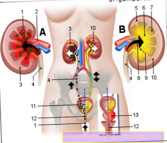
Pelvic inflammation
(bacterial infection of the
Renal pelvis)
Pyelonephritis
You can find an overview of all Dr-Gumpert images at: medical illustrations
$config[ads_text2] not found$config[ads_text3] not found
The diagnosis of enlarged kidneys is often made using an ultrasound. Here, the position and size are determined. In addition, it is possible to see the flow of urine and possibly urinary stones. Furthermore, possible space demands inside or outside the kidney can be detected.
X-rays can show kidney or urinary stones as the cause of the enlarged kidney.
Computed tomography can also provide information about the reason.
With an MRI of the kidney (nuclear spin tomography) vascularization can be seen possible neovascularization of a tumor or a thrombosis of the renal artery.
Laboratory tests of the blood and urine can also provide information about the causes. A tissue sample can also be taken from the kidney (kidney biopsy) to clarify unclear inflammations.
The possible accompanying symptoms of kidney enlargement can be as diverse as their causes. Decreased amount of urine, bloody urine and Painful urination can indicate a urinary stone disorder.
To step Fever, chills, and water retention (Edema) in the legs or on the eyelids, this can be due to a Inflammation of the kidney indicate that due to the loss of protein due to the impaired filter function of the kidneys, foamy urine can also occur.
At a Diabetes insipidus (Water clock) the daily amount of urine can be increased and associated with a greatly increased feeling of thirst and the corresponding amount of drinking.
In some cases, the kidney enlargement can be felt under the skin. If the cause is an abscess, it is possible to find local reddening of the skin over the kidney.
There are also a number of them unspecific symptoms such as exhaustion and a general feeling of illness, which can be associated with reduced physical performance.
Kidney pain Depending on the cause, they can be acute colic or persistent. A flank pain is often described as well as a Pounding pain over the kidneyswhich can be triggered by tapping the back with your fist at kidney level.
Pain in the groin region can result from the pressure of the enlarged kidney on the adjacent nerves. The kidney capsule is sensitive to pain and can hardly be stretched. If there is inflammation, the resulting swelling and the limited space can cause severe pain.
First is an adequate one Pain relief the most important measure through potent pain medication. The further treatment strategies depend on the cause.
Blocks a Urinary stone the ureters and prevents the urine from flowing out, stones smaller than 5 millimeters and a course without complications can be awaited for the stone to pass spontaneously. The person affected should move around a lot and drink a lot of fluids. The doctor can also prescribe medication that promotes stone loss. The urine should then be passed through a sieve to check for possible stone loss.
Read about this: Therapy of kidney stones
To Prevention of urinary stones In general, a generous intake of at least 2.5 liters per day should be taken. In addition, a balanced, low-sodium diet is recommended.
Read about this: This is the best way to prevent kidney stones
The duration of a kidney enlargement again depends on the cause. If, for example, the urinary calculus goes away in the case of a stone disease, the kidney can return to its original size relatively soon. In this case, it also depends on the severity of the clinical picture.
If the mother's kidney enlarges during pregnancy due to constricting anatomical conditions, a rapid normalization of the size can be observed after the birth of the child.
Physiologically it is right kidney smaller and laid out lighter than the left one.
If a bacterial inflammation is the cause of the enlargement of the kidney, it is not necessarily the case that the second kidney is also affected. However, this can be the case, for example, with an ascending urinary tract infection.
$config[ads_text1] not found
With the multitude of pregnant women An enlargement of the renal pelvis can be observed. Here, due to the increase in size of the uterus, the right ureter is more frequently relocated and the calyx system of the calyx system increases in size temporarily right kidney.
With the heritable polycystic kidney disease there is a participation both kidneys. Solitary kidney cysts can occur on one or both sides; these are often symptom-free and are often discovered as an incidental finding.
In babies, an enlarged kidney can occur in connection with a narrowing of the ureter immediately at the exit from the kidney (Ureteral obstruction). The cause of this can be a disturbance of the smooth muscles, which is associated with impaired peristalsis in a segment of the ureter and thus hinders the outflow of urine from the kidney. On the other hand, incorrectly placed vessels can cause a narrowing of the ureter in the upper part, but also a position deviating from the norm or a changed shape of the kidney.
Here it is important that Monitor renal function closely to initiate any therapeutic measures. Parents should also pay attention to the amount of urine their baby has in the diaper.
The malposition in a part of the urinary bladder can lead to a so-called in the fetus Vesicoureteral reflux to lead. A possible malposition is at the point of the urinary bladder where the ureters open. Another possibility of incorrect investment can be a double ureter be.
In vesicoureteral reflux, the urine is transported back into the ureters from the bladder due to neuronal innervation. The complication of this reflux can range in to frequent inflammation with fever Urosepsis, a serious clinical picture. In addition, severe kidney dysfunction, terminal kidney failure, can occur.
The therapy of these infections should be controlled with medication. The prognosis is associated with a high spontaneous healing rate, but repeated and uncontrollable infections should make the child prick up his ears. In these cases, surgical therapy can result in a significant improvement in the symptoms.
The renal pelvis is the urine collection basin, after which the functional units of the kidney have filtered and concentrated the urine. The shape of the renal pelvis depends on the structure of the calyxes which open into the pelvis. In the case of particularly narrow goblets (dendritic type) there is not much space for enlargement, so that space is limited in the event of urine accumulation.
In the case of hydronephrosis (pent-up urine) as a result of a stone in the ureter, the renal pelvis can be enlarged. In some cases, a large urinary stone can also line the entire surface of the renal pelvis and limit its flexibility.