Rib cartilage, also called cartilago costalis, is the connection between the ribs and the breastbone (sternum).
The costal cartilage thus forms the last part of the ribs, which are connected to the sternum via these.
The costal cartilage thus forms part of the anterior human rib cage.
The costal cartilage is a hyaline cartilage that is both compressive and flexurally elastic compared to the bony ribs and bony sternum.
In early adulthood, the cartilage gradually begins to calcify and later also to ossify, which reduces the elasticity of the chest more and more with age.

The costal cartilage is the connection between the ribs and the breastbone (sternum).
The costal cartilage forms the last 3 - 9 cm of the ribs that are connected to the sternum.
The cartilages of the six upper ribs are attached to the sternum by ligaments, the sternocostal ligaments.
Ribs six and seven are also attached to the lower part of the sternum, the xiphoid process, by a ligament, the costoxiphoid ligament.
$config[ads_text1] not found
The costal cartilage forms part of the anterior rib cage and is partially palpable from the outside, as the boundaries between the bony ribs and the cartilage tissue are often slightly thickened.
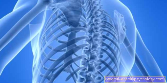
The ribs, together with the spine and sternum, form the bony framework of the chest.
The ribs arise from the spine and run along the lungs forwards, mostly to the sternum.
When it comes to ribs, a distinction is made between so-called “true ribs”, “false ribs” and “free ribs”.
A person has a total of twelve ribs.
When viewed from above, the “true ribs” are the first seven ribs and are directly connected to the sternum via the costal cartilage.
The following three ribs, i.e. ribs eight to ten, are only indirectly connected to the sternum via the so-called articulatio interchondrales with the costal cartilage of the upper seven ribs.
The last two lower ribs have very short or no costal cartilages and are therefore not connected to the sternum.
The chest surrounds the two lungs and is only separated from them by the pleural cavity.
The pleural cavity is a very narrow body cavity that is filled with 5-10 milliliters of serous fluid.
This reduces the friction between the rib cage and the lungs.
Since the front parts of the ribs are lined with hyaline cartilage, the rib cage has slightly elastic properties.
These elastic properties are crucial for the freedom of movement of the chest.
The essential function of the costal cartilage is to ensure the elasticity of the rib cage.
Since the costal cartilage also forms part of the chest, it also serves to protect the underlying lungs and heart.
The costal cartilage is made up of hyaline cartilage.
The hyaline cartilage is widespread in the body and can often be found in joints.
Compared to bony structures, cartilage is elastic in compression and bending.
If you exert pressure on the cartilage, it can initially withstand the pressure by bending.
When the pressure is removed, the cartilage quickly returns to its original position thanks to its elasticity.
Bony structures show these properties only to a very small extent - they would break quickly if the same pressure were exerted.
For this reason, the costal cartilage is essential for the elasticity of the rib cage, which is particularly important when breathing in and out.
$config[ads_text3] not found
In addition, the costal cartilage of the second to sixth ribs is the starting point for the musculus thoracicus transversus, which pulls from the inside to the inside of the sternum.
The muscle mentioned supports the exhalation.
Rib cartilage pain is usually caused by inflammation or damage to the cartilage.
The symptoms occur in the anterior costal arch, usually at the level of the fourth to seventh rib.
Tietze syndrome is a well-known but very rare syndrome that is associated with damage to the cartilage.
Tietze syndrome results in pain and swelling in the anterior chest area, mainly localized on the costal cartilage.
The pain usually occurs suddenly and can also radiate into the arms and shoulders, which is why they have symptoms similar to angina pectoris and are often confused with them.
The pain increases with deep inhalation.
In order to rule out the differential diagnosis of angina pectoris, the chest is scanned at the points where the costal cartilage is located.
The cause of a Tietze syndrome is usually exceptional stress (e.g. heavy lifting / pulling) or trauma.
Tietze syndrome is usually only treated with painkillers and heals itself over time.
An alternative explanation for pain in the costal cartilage is its inflammation, also known as costochondritis.
Even if the costal cartilage is inflamed, the pain can be transmitted to the back or abdomen.
The pain is particularly felt when inhaling deeply, coughing, sneezing or laughing.
As with Tietze syndrome, the cause can be exceptional stress.
But infections of the respiratory tract can also spread to the cartilage and lead to its inflammation.
Treatment is also not usual for inflammation of the cartilage.
The only way to treat pain is to prescribe pain medication. However, vigorous physical work should be absolutely avoided during costal cartilage inflammation.
Generally speaking, damage to or inflammation of the costal cartilage is harmless.
However, the symptoms that one exhibits are very similar to those of a heart attack, which is why a comprehensive examination should be made by a doctor in any case if pain occurs.
Are you interested in this topic? Read more about this under: Rib pain - these are the causes
The costal cartilage is the connection between the ribs and the sternum.
A rupture of the costal cartilage is extremely rare, but it is associated with severe pain.
The costal cartilage consists of hyaline cartilage and is usually elastic in compression and bending.
This means that if the pressure exerted on the costal cartilage is light to medium, it will initially bend slightly before it breaks if the pressure is too strong.
Since the costal cartilage increasingly begins to calcify as a result in early adulthood, its elasticity also increasingly decreases with age.
This means that the risk of a costal cartilage rupture increases with age.
Rib cartilage fractures often occur as a result of resuscitation or severe trauma.
Because the costal cartilage creates the connection between the rib and the sternum, fractures of the rib cartilage tend to occur in connection with fractures of the sternum (sternum) or fractures of the rib.
Alternatively, rib cartilage tears can rarely occur.
The costal cartilage is attached to the sternum by ligaments, the ligamenti sternocostales radiatum.
These ligaments can tear as a result of strong pressure or extreme expansion of the chest.
The connection between the ribs and the sternum is lost.
Particularly when you breathe in deeply, with the front part of the chest exerting strong movement, severe pain can occur.
There is no clear treatment for both the fracture of the costal cartilage and the tear of the costal cartilage.
Most of the time, only pain medication is prescribed.
In addition, intensive rest and rest are recommended for rapid improvement of the symptoms.
The swelling of the costal cartilage is mostly due to the so-called Tietze syndrome.
Alternatively, swelling can also occur due to polychondritis.
Tietze syndrome is a rarely occurring inflammation in the area on the side of the sternum.
The inflammation is usually localized at the level of the upper ribs at the transition to the sternum, i.e. the costal cartilage.
There is severe pain in the area of the sternum, which can radiate into the left arm and back.
Therefore, the differential diagnosis of myocardial infarction should definitely be clarified.
There is also a risk that the inflammation of the costal cartilage can spread to nearby organs, such as the heart and lungs.
The X-ray does not show any abnormalities in Tietze syndrome.
Alternative differential diagnoses are other inflammations in the area of the rib skeleton, which, however, in comparison to Tietze syndrome, do not show any swelling and are mostly located on the side.
Treatment for Tietze syndrome consists solely of treating the symptoms.
For this reason, pain killers can usually only be used.
A chronic, long-lasting cartilage disease is generally referred to as polychondritis.
The cause of this are autoimmune inflammatory processes in which the cartilage tissue perishes during a lengthy process.
Since the costal cartilage consists of hyaline cartilage, it is also affected by polychondritis, which can lead to swelling.
However, in polychondritis the swelling of the costal cartilage does not occur in isolation, but rather a cartilage swelling throughout the body, which can be used to differentiate it from Tietze syndrome.
Inflammation of the rib cartilage is the inflammation of the joint that connects the ribs to the sternum.
The joint is also known as the sterno-costal joint.
The inflammation is usually accompanied by severe pain and is also called costochondritis.
Typical symptoms of costal cartilage inflammation are swelling and pain in the upper chest area on the side of the sternum.
The pain can be very severe and long-lasting and can usually only be treated with painkillers until the inflammation subsides on its own.
Costochondritis occurs more frequently in young adults between the ages of 20 and 40.
The cartilage inflammation can be triggered by various causes.
One possible cause can be severe trauma to the chest area.
Alternative triggers are transmitted infections, rheumatism or the consequences of Tietze syndrome.
The pain during costal cartilage inflammation can radiate to the back and arm.
They also occur more frequently when you breathe deeply, cough, sneeze or laugh.
Inflammation is usually harmless unless it spreads to surrounding organs such as the heart and lungs.
The cracking of the costal cartilage can usually be traced back to the joint that connects the ribs and sternum, the articulatio sternocostales.
This can be found between the first six ribs and the sternum.
It can easily block or tilt.
Removing this blockage or tilting can then cause the joint to crack, but is usually absolutely harmless.
In addition, if the ribs or the costal cartilage break, the connection between them and the sternum can be broken.
Moving this - for example when inhaling - can cause cracking in the area of the ribs or the sternum.
If this type of cracking costal cartilage occurs, pain is definitely associated with it, while there is usually no pain when the canting of the sterno-costal joint is lifted.
Rather, the cracking can relieve pain as a result of unblocking a block.
How to properly solve a rib block, read here: This is the correct way to solve a rib block
The costal cartilage consists of a hyaline cartilage.
This is characterized by its compressive and flexural elasticity.
Beginning in early adulthood, however, the cartilage begins to calcify, which leads to increased elasticity restrictions.
The reason for the calcification of the ribs are metabolic processes that occur more strongly in men than in women.
Calcium accumulates in the cartilage, which stiffens it.
Read more on the subject at: Chondrocalcinosis - Causes & Symptoms