
The spinal canal is also called the spinal canal or spinal canal. It is formed by the foramina vertebralis of the vertebral bodies of the cervical, thoracic and lumbar spine as well as the sacrum and in it is the spinal cord, which is protected by meninges. The canal is drawn to the front and to the side by the vertebral arches and the ligaments connecting them (Ligamenta flava) bounded and towards the rear by the vertebral bodies and the posterior longitudinal ligament (posterior longitudinal ligament).
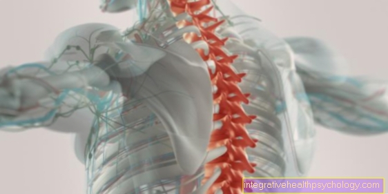
The Spinal cord is also attached by ligaments in the spinal canal and the spinal fluid is found around the spinal cord (Cerebrospinal fluid). In healthy adults, the spinal cord ends approximately at the level of the lumbar vertebrae L1 / 2. Underneath, the nerves pull as a bundle, the so-called cauda equina (ponytail). Pull at the level of each vertebral body two spinal nerves of the spinal cord laterally from the spinal canal and supply the corresponding segments.
$config[ads_text1] not found
The spinal cord is like the brain surrounded by the 3 meninges. The outer skin is the dura mater. It has two leaves. The outer leaf lies directly on the vertebrae. In the space between the two leaves is the front and back of the spinal canal venous plexus (Plexus venosus vertebralis internus anterior and posterior). This room is also called Peri- or epidural space. They also run in it Arteries to supply the spinal cord, which form the anterior spinal artery. The inner leaf of the dura mater rests on the arachnoid and forms so-called dural sagging, which continues a short distance with the spinal nerves.
Under the dura mater lies the Arachnoidthat, like that, pulls a piece with the spinal nerves. Both also pull down to the end of the spinal canal and, like the spinal cord, don't end before that. Between the arachnoid and the pia mater lying directly on the spinal cord lies the Subarachnoid space. He's for that CSF puncture important below L 1/2.
The most important function of the spinal canal is Protection of the spinal cord. The spinal cord is the connection from the brain to all organs, muscles, etc. and when this does not work properly it leads to paralysis, organ failure or other restrictions, so protecting it is very important. A particularly dreaded complication of spinal cord injury is Paraplegia. Depending on the severity of the injury, this can also be fatal.
$config[ads_text2] not foundThis is guaranteed by 3 different components of the spinal canal. On the one hand, the ligaments and the skeleton form a stable, difficult to deform channel in which the spinal cord lies. External shocks are also cushioned by the venous plexus and the fatty tissue in the epidural space. They represent a cushion, so to speak. Finally, the spinal cord is protected by the liquor. This gives the spinal cord a certain mobility but at the same time prevents it from hitting the bone.
In addition, the spinal canal enables safe exit of the spinal nerves from the spinal cord and thus ensures the supply of the periphery.
Another function of the spinal canal is that Formation of the liquor space. The liquor is not only a protective cushion for the spinal cord, but also serves the metabolism of the nerve cells. The CSF puncture, which is also taken from the spinal canal, is an important diagnostic criterion. It takes place below the lumbar vertebra L1 / 2 at the level of the cauda equina in order to protect the spinal cord as much as possible.
With the CSF puncture, various inflammatory diseases of the brain can be diagnosed by determining the number of cells, the protein content, the antibodies and the like. You can do that Subarachnoid hemorrhage, different tumors, one meningitis or Encephalitis and also multiple sclerosis diagnose.
$config[ads_text3] not found
Spinal canal stenosis is a narrowing of the spinal canal, which can have various causes. This narrowing can lead to pressure on the spinal cord and the vessels in the canal, which can lead to back pain and even nerve damage.
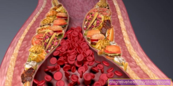
$config[ads_text2] not foundThe pain mostly depends on the degree of constriction and posture. The pain usually gets worse when standing with an upright back, while it tends to subside when the back is bent. The most common cause of spinal stenosis is aging. The spine is changed by osteoporosis and bone remodeling processes, especially in the neck and lumbar area. The spaces between the vertebrae become smaller and smaller and the ligaments lose tension. They wear out, so to speak. Due to these two phenomena, the eddies can shift against each other. If the back muscles are not well developed, new bone formation occurs (Osteophytes), which can then narrow the spinal canal. This can narrow the canal. Usually it is a lumbar spinal stenosis.
Other causes can include congenital constrictions, spinal injuries, herniated discs and bone diseases.
The diagnosis is usually made by a CT or MRI. The course of the disease depends on the severity of the narrowing of the canal, but most of the time the disease progresses very slowly. In mild forms, no intervention is necessary as it is usually symptom-free.
In very severe cases, surgery may also be indicated to relieve the patient's pain. The standard therapy is mostly through pain medication and physiotherapy.
Read more on the subject at:
$config[ads_text4] not found
The symptoms are usually back pain, restricted mobility and muscle tension, especially in the lumbar spine. If the constriction increases, there are also sensory disturbances, paresthesia, weakness, as well as bladder and sexual disorders due to the increased constriction of the spinal cord.
Read more on the subject at: Spinal canal stenosis in the lumbar spine
Tumors in the spinal canal usually arise when spinal tumors grow into the canal. So they do not have their origin in the spinal canal, but in the spine. Spinal tumors can either primary be, that is, they arise directly in the bones of the spine, or they are secondarythat means it is Metastases from other tumors.
There are benign and malignant tumorsthat can form in the spine. To the benign Tumors include osteoid osteomas, osteoblastomas, hemangiomas, fibrous histiocytomas, aneurysmal bone cysts, and eosinophilic granulomas. They mostly make up only noticeable through pain and only very rarely through neurological deficits. Most benign tumors are incidental findings. They are usually only treated if the spinal canal is also affected.
Vicious Tumors are for example Ewing's sarcoma, osteosarcoma and chondrosarcoma. Due to their invasive growth, these represent a great danger to the spinal cord and therefore have to be operated on if possible and chemotherapy and radiation therapy are usually also necessary. Metastases in the area of the spine are also quite common and can lead to damage to the spinal cord and even paraplegia due to vertebral deformities and the tumor growing into the spinal canal.Here too, rapid and intensive therapy is necessary.
Probably the most important type of syringe into the spinal canal is peridural infiltration. Medicines are slowly injected into the epidural space (space between the two sheets of the outer meninges) of the spinal cord. The active ingredients are Local anesthetics and glucocorticoids. Local anesthetics are used for local anesthesia, for example during surgery or for pain relief. Glucocoticoids, such as cortisone, are used to treat inflammation. In combination, they are used to treat various orthopedic diseases, for example nerve root syndrome (damage to the spinal nerve due to narrowing of the spinal canal), the disc prolapse or spinal stenosis.
$config[ads_text1] not foundThe Spinal anesthesia is a form of local anesthesia for operations in the area of the lower abdomen, groin, perineum and legs. It is best known for its use in caesarean sections. Here the active ingredient is injected into the subdural space and is therefore effective very quickly. The Epidural anesthesia (PDA) is especially important for pain relief and is mainly used in obstetrics to relieve pain during labor. The PDA is injected into the epidural space and therefore works somewhat more slowly than the spinal anesthesia.


.jpg "Bioelectrical Impedance Analysis (BIA)")


