
The human eye has two types of photoreceptors that enable us to see. On the one hand there are the rod receptors and on the other hand the cone receptors, which are further subdivided: blue, green and red receptors. These photoreceptors represent a layer of the retina and send a signal to the transmitting cells linked to them if they detect an incidence of light. The cones are used for photopic vision (color vision and vision by day) and the rods, on the other hand, for scotopic vision (perception in the dark).
More on this subject: How does vision work?
The human retina, too retina called, is a total of 200 µm thick and consists of different cell layers. The pigment epithelial cells, which are very important for the metabolism, lie on the outside retina by absorbing and breaking down dead photoreceptors and also secreted cell components that arise during the visual process.
$config[ads_text1] not found
The actual photoreceptors, which are separated into rods and cones, now follow inward. Both have in common that they have an outer limb, which points towards the pigment epithelium and also has contact with it. This is followed by a thin cilium, through which the outer link and inner link are connected to one another. In the case of the rods, the outer link is a layer of membrane disks, similar to a stack of coins. In the case of the tenons, however, the outer link consists of membrane folds so that the outer link looks like a kind of hair comb in a longitudinal section, with the teeth representing the individual folds.
The cell membrane of the outer limb contains the visual pigment of the photoreceptors. The color of the cones is called rhodopsin and consists of a glycoprotein opsin and 11-cis retinal, a modification of vitamin A1. The visual pigments of the cones differ from rhodopsin and from each other by different forms of opsin, but also have the retinal. The visual pigment in the membrane disks and membrane folds is consumed by the visual process and has to be regenerated. The membrane disks and folds are always newly formed. They migrate from the inner member to the outer member and are ultimately released and absorbed and broken down by the pigment epithelium. A malfunction of the pigment epithelium causes a deposition of cell debris and visual pigment, as occurs for example in the disease of the Retinitis pigmentosa is.
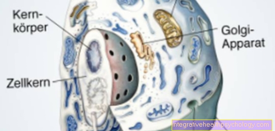
$config[ads_text2] not foundThe inner member is the actual cell body of the photoreceptors and contains the cell nucleus and the cell organelles. Important processes take place here, such as the reading of DNA, the production of proteins or cell messenger substances; in the case of photoreceptors, glutamate is the messenger substance.
The inner limb is thin and has a so-called receptor foot at the end, through which the cell is connected to so-called bipolar cells (forwarding cells). Transmitter vesicles with the messenger substance glutamate are stored in the receptor base. This is used to transmit signals to the bipolar cells.
A special feature of the photoreceptors is that when it is dark, transmitter substance is permanently released, whereby the release decreases when the light falls. So it is not like with other perception cells that a stimulus leads to an increased release of transmitters.
There are rod and cone bipolar cells, which in turn are interconnected with ganglion cells, which make up the ganglion cell layer and whose cell processes ultimately together form the optic nerve. There is also a complex horizontal interconnection of the cells of the retinawhich is realized by horizontal cells and amacrine cells.
The retina is stabilized by so-called Müller cells, the glial cells of the retinawhich span the entire retina and act as a framework.
$config[ads_text3] not found
The photoreceptors of the human eye are used to detect incident light. The eye is sensitive to light rays with wavelengths between 400 - 750 nm. This corresponds to the colors from blue to green to red. Light rays below this spectrum are referred to as ultraviolet and above as infrared. Both are no longer visible to the human eye and can even damage the eye and cause lens opacity.
$config[ads_text2] not foundMore on this topic: Cataract
The cones are responsible for color vision and require more light to emit signals. In order to realize color vision, there are three types of cones, each of which is responsible for a different wavelength of visible light and has its absorption maximum at these wavelengths. The photopigments, the opsins of the visual pigment of the cones, therefore differ and form 3 subgroups: the blue cones with an absorption maximum (AM) of 420 nm, the green cones with an AM of 535 nm and the red cones with an AM of 565 nm. If light of this wavelength spectrum hits the receptors, the signal is passed on.
More on this topic: Examination of color vision
Meanwhile, the rods are particularly sensitive to the incidence of light and are therefore used to detect even very little light, especially in the dark. It is only differentiated between light and dark, but not in terms of color. The visual pigment of the rod cells, also called rhodopsin, has an absorption maximum at a wavelength of 500 nm.
As already described, the cone receptors are used for daytime vision. Through the three types of cones (blue, red and green) and a process of additive color mixing, the colors we see can be seen. This process differs from physical, subtractive color mixing, which is the case, for example, when mixing painters' colors.
$config[ads_text4] not found
In addition, the cones, especially in the viewing pit - the place of sharpest vision - also enable sharp vision with a high resolution. This is also due in particular to their neural interconnection. Fewer cones lead to a respective ganglion neuron than with the rods; the resolution is therefore better than with the chopsticks. In the Fovea centralis there is even a 1: 1 forwarding.
The rods, on the other hand, have a maximum with an absorption maximum of 500 nm, which is right in the middle of the visible light range. So you react to light from a wide spectrum. However, since they only have the rhodopsin, they cannot separate light of different wavelengths. However, their great advantage is that they are more sensitive than the cones. Significantly less incidence of light is also sufficient to reach the reaction threshold for the rods. They are therefore used to see in the dark when the human eye is color blind. The resolution, however, is much worse than with the cones. More rods converge, i.e. converge, lead to a ganglion neuron. This means that regardless of which rod from the bandage is excited, the ganglion neuron is activated. So there is no such good spatial separation possible as with the tenons.
It is interesting to note that the rod assemblies are also the sensors for the so-called magnocellular system, which is responsible for movement and contour perception.
In addition, one or the other may have already noticed that stars are not in the focus of the field of vision at night, but rather on the edge.This is because the focus projects onto the pit of view, but it has no chopsticks. These lie around them, so you can see the stars around the focus of the gaze center.
Due to their different tasks, the cones and rods in the eye are also distributed differently in terms of their density. The cones are used for sharp vision with color differentiation during the day. You are therefore at the center of the retina most common (yellow spot - Macula lutea) and in the central pit (Fovea centralis) are the only receptors present (no rods). The viewing pit is the place of sharpest vision and specializes in daylight. The rods have their maximum density parafoveal, i.e. around the central pit of vision. In the periphery the density of the photoreceptors decreases rapidly, whereby in the more distant parts almost only rods are present.
$config[ads_text1] not foundCones and chopsticks share the blueprint to some extent, but then vary. In general, chopsticks are slightly longer than cones.
Rod photoreceptors average about 50 µm in length and about 3 µm in diameter at the most densely packed locations, i.e. for rods, the parafoveal region.
The cone photoreceptors are somewhat shorter than the rods and have a diameter of 2 µm in the fovea centralis, the so-called pit of vision, in the region with the highest density.
The human eye has an overwhelming number of photoreceptors. One eye alone has around 120 million rod receptors for scotopic vision (in the dark), while there are around 6 million cone receptors for day vision.
Both receptors converge their signals on to about a million ganglion cells, whereby the axons (cell extensions) of these ganglion cells make up the optic nerve (nervus opticus) as a bundle and pull into the brain so that the signals can be centrally processed there.
More information can be found here: Visual center
As already described, rods and cones have slight differences in structure, which are, however, not serious. Much more important is their different function.
Rods are much more sensitive to light and can therefore detect even small incidence of light, but only differentiate between light and dark. In addition, they are slightly thicker than the cones and are passed on in a converging manner, so that their resolving power is lower.
Cones, on the other hand, require more incidence of light, but can enable color vision through their three sub-forms. Due to their smaller diameter and the less converging transmission, up to 1: 1 transmission in the fovea centralis, they have an excellent resolution, which can only be used during the day.
The Macula lutea, also known as the yellow point, is the place on the retina with which people primarily see. The name was given by the yellowish coloration of this point in the fundus of the eye. The yellow spot is the place of the retina with most photoreceptors. Except for Macula there are almost only rods left that are supposed to differentiate between light and dark.
The Macula centrally still contains the so-called viewing pit, Fovea centralis. This is the point of sharpest vision. The viewing pit contains only cones in their maximum packing density, whose signals are transmitted 1: 1, so that the resolution is best here.
Dystrophies, pathological changes in body tissue that cause the retina are usually genetically anchored, i.e. they can either be inherited from the parents or acquired through a new mutation. Some medications can cause symptoms similar to retinal dystrophy. The diseases have in common that symptoms only appear in the course of life and they have a chronic, but progressive course. The course of dystrophies can vary greatly from disease to disease, but it can also vary greatly within one disease. The course can even vary within an affected family, so that no general statements can be made. In some diseases, however, it can progress to blindness.
$config[ads_text2] not found
Depending on the disease, visual acuity can decrease very quickly or gradually deteriorate over several years. The symptoms, whether the central field of vision changes first or the loss of field of vision progresses from the outside to the inside, is variable due to the illness.
Diagnosing retinal dystrophy can be difficult at first. However, there are numerous diagnostic procedures that can make a diagnosis possible; here is a small selection:
Unfortunately, it is currently the case that no causal or preventive therapy is known for most of the genetically caused dystrophic diseases. However, a lot of research is currently being carried out in the area of genetic engineering, with these therapies currently only in the study phase.
The human visual pigment consists of a glycoprotein called opsin and the so-called 11-cis-retinal, which is a chemical modification of vitamin A1. This also explains the importance of vitamin A for visual acuity. In the case of severe deficiency symptoms, night blindness and in extreme cases blindness can occur.
Together with the 11-cis retinal, the opsin produced by the body itself, which exists in various forms for rods and the three cone types (“cone opsine”), is built into the cell membrane. When exposed to light, the complex changes: the 11-cis retinal changes to the all-trans retinal and the opsin is also changed. Metarhodopsin II, for example, is produced in the rods, which sets a signal cascade in motion and reports the incidence of light.
Red-green weakness or blindness is a malfunction of color vision that is congenital and inherited X-linked with incomplete penetrance. However, it can also be that it is a new mutation and therefore none of the parents has this genetic defect. Since men only have one X chromosome, they are much more likely to get the disease and up to 10% of the male population is affected. However, only 0.5% of women are affected, as they can compensate for a defective X chromosome with a healthy second one.
The red-green weakness is based on the fact that a genetic mutation has taken place for the visual protein opsin in either its green or red isoform. This changes the wavelength to which the opsin is sensitive and therefore red and green tones cannot be sufficiently distinguished. The mutation occurs more frequently in the opsin for green vision.
There is also the possibility that color vision for one of the colors is completely absent, for example if the coding gene is no longer present. A red weakness or blindness is called Protanomaly or. Protanopia (for green: Deuteranomaly or. Deuteranopia).
A special form is the blue cone monochromatism, i.e. only the blue cones and blue vision work; Red and green cannot then be separated either.
Read more on the topic:


.jpg "Extrasystoles (palpitations)")


