Scintigraphy
definition
Scintigraphy is an imaging process that plays a crucial role in nuclear medicine diagnostics.
In order to generate an image, a so-called scintigram, the patient is given radioactive substances.
These emit radiation and can then be detected in the corresponding organ or tissue using a gamma camera.

execution
With the help of a radioactive substance, tissue or organs can be specifically examined.
To do this, the patient is injected with radioactive material.
The patient can either receive the radioactive substances directly by injection or they can be administered orally as tablets.
Depending on which tissue or organ you want to represent, different substances are suitable.
For example, there are substances that accumulate particularly well in bone tissue.
This substance, which is specific for a tissue, is called a tracer. For example, there is a radioactive iodine particle for examining the thyroid gland or 99mTc-iminodiacetic acid for examining the hepatobiliary function (i.e. the functionality or liver and gallbladder).
In the case of bones, this is usually the technetium isotope 99mTc.
This is deposited in the bone and remains there. The particle now emits gamma rays from the bone.
These gamma rays can be detected using a camera. A color-visualized image now appears on the computer.
The more frequently the particle emits so-called flashes of light, i.e. gamma rays, the blacker the spot in the image appears.
In the case of a color image, the color blue stands for a low level of activity of the radioactive particles in the tissue, in the case of red color the radioactive particles are very active.
In this way, the radioactively marked particles can be used to find out how active the tissue is. If areas of the thyroid gland light up blue on a scintigram, you can be sure that this part of the thyroid gland is no longer properly active for some reason.
At the same time you can recognize a focus of inflammation by the red glowing color.
If there is an inflammation in an organ, the metabolism is much more intense. There is increased blood flow and activity is increased.
This can be seen very well on the basis of the scintigram and thus an accurate diagnosis can be made.
Duration of a scintigraphy
A scintigraphy can usually be performed very quickly.
Depending on the type of tissue to be examined, the examination takes 10 minutes to an hour.
However, the length of the preparation time is important.
Since during an examination of the thyroid gland drugs against an overactive thyroid or underactive thyroid have to be discontinued, these "Preparations" a day.
It is also important to note that some radionucleotides take a long time to be absorbed by the corresponding tissue.
It is therefore possible that after the administration of the radioactive substances, the examination can take place after 10 minutes or only after several days.
It is also possible that one measurement is not sufficient and that a control measurement must take place.
Working principle
The creation of the scintigraphic image (scintigram) is based in principle on the Detection of radioactive pharmaceuticals. This so-called Tracer substance (Radionuclide) is bound to a certain carrier that is specific for the tissue that is to be displayed and preferably accumulates there (e.g. iodine to display the thyroid; bisphosphates to display bones).
The injected radionuclide, as unstable isotope, has the property of emitting radiation (preferably? -radiation) when it decays, which is then triggered by a Gamma camera can be recorded. In most cases the technetium isotope 99mTc is used as a radioactive nuclide.
The ones captured by the gamma camera gamma rays are then by a so-called, located in the camera Scintillation crystal in Flashes of light and further converted into electrical signals in the course. This electrical signals are then as Blackening in the Scintigram visible. The degree of blackening depends on the frequency of the radiation, i.e. on the Amount of enriched radioactive substance in the respective organ / tissue. The more a tissue accumulates, the darker it appears in the image.
Forms of scintigraphy
In the Scintigraphy two types can be distinguished in imaging.
On the one hand, the static scintigraphy can be used in which the distribution in the respective organ / tissue is detected only at a predetermined point in time after the injection of the radiopharmaceutical.
On the other hand, however, a dynamic scintigraphy be carried out, with both the Flooding as well as the flooding process of the radiopharmaceutical is represented in the organ / tissue. This makes an accurate Depiction of blood flow in certain regions as well as answering certain questions such as the function of the kidneys or the Elimination ability of the liver possible.
With the one mentioned above SPECT procedure, one combination out Scintigraphy and Computed tomography, in addition to three-dimensional imaging, static and dynamic components can also be recorded.
Frequency distribution
Since scintigraphy sheds light on most of the Organ functions can give, it is very suitable as an imaging procedure.
Besides, the Radiation exposure lower than in comparison with X-rays. That is why around 60,000 scintigraphies are made every week in Germany. Most of them are used to examine the thyroid gland.
diagnosis
Various diagnoses can be made with the aid of scintigraphy.
The most common indication for scintigraphy is an examination of the thyroid gland. With the help of the radioactively labeled substances, for example, one can determine an overfunction.
In this case, after the tracer has been injected, the tissue would be unusually red, i.e. unusually active.
However, one can also have a cyst or a malignant tumor (Carcinoma) detect.
In these cases, too, the tissue would be more metabolically active because a tumor needs a lot of energy.
On the other hand, inflammation or metastases can be seen on the skeleton. An examination of the lungs, heart or kidneys is a rare indication for a scintigraphy.
With the help of a scintigram, however, a diagnosis of a possible pulmonary embolism, a narrowing of the coronary arteries (Coronary arteries) or narrowing of the renal arteries.
In addition to establishing a diagnosis, scintigraphy can also be used as a therapy control.
For example, one examines the heart to see whether the coronary arteries have expanded after appropriate therapy (Myocardial scintigraphy).
Or you can do a ventilation scintigraphy to check that your lungs are properly ventilated while you breathe.
Therefore, indications for a scintigraphy are always the verification of a diagnosis.
For example, if the doctor suspects that the patient may suffer from an overactive thyroid based on the anamnesis, i.e. the doctor-patient consultation, this initial diagnosis can be confirmed with the help of scintigraphy.
In order to be able to perform a scintigraphy, the patient must adhere to certain rules so that the diagnosis is also safe and reliable.
For example, if a patient is taking medication for an overactive thyroid, they must stop taking them before treatment.
If the patient does not stop taking the medication, scintigraphy cannot be used to make an exact judgment, as the thyroid activity is falsified by taking the medication.
When examining the heart, the patient should appear for the examination on an empty stomach, i.e. he must not have drunk or eaten for several hours before the examination.
execution
Before the start of the Scintigraphy are commonly no major preparations necessary. However, depending on which organ / tissue is to be examined, certain specifications can be made so that the medication cannot always be continued or an empty state (especially when examining the gastrointestinal tract) must be observed.
At the beginning of the scintigraphic examination, the patient receives this radioactive agents through the arm vein injected into the bloodstream (usually through the vein in the crook of the elbow). Afterwards, depending on the radiopharmaceutical used, you have to wait for different lengths of time until the radioactive substance has distributed in the body and accumulated in the desired tissues / organs (there are usually waiting times between a few minutes and 1-3 hours).
Since that injected Radiopharmaceutical usually excreted through the kidneys care should be taken that the The patient drinks a lot of fluids while waiting and visits the toilet several times to find the radioactive substance accumulated in the bladder to prevent. Due to the faster excretion, this reduces the radiation exposure on the one hand and enables one on the other better resolution and quality of the recordings.
When creating the scintigram, the patient sits or lies in prone or supine position under the detecting gamma camera, which is usually a predominantly open camera system represents (not a pipe system like a MRI/CT).
The Recording time varies also and depends on the organ to be recorded and the respective question: the imaging of the thyroid gland as a relatively small organ takes about 5 minutes on average, the display of the bones or the entire skeleton, however, about 20-40 minutes to 1 hour. The patient should lie / sit as quietly as possible during the entire examination in order to prevent “blurring” of the image and to enable the most precise and sharp scintigram possible.
Duration of a scintigraphy
How long a scintigraphy takes depends on the organ examined and the radioactive substance used. On the one hand, the time period from injection to uptake and distribution in the target organ varies. On the other hand, the radioactive particles decay at different speeds. In addition, the period of time required for recording with the camera is different for each type of scintigraphy.
It follows that a thyroid scintigraphy is usually finished after 30 minutes. Allow 30 to 60 minutes for the lungs and kidneys.In contrast, bone and heart scintigraphy, in particular, can take significantly more time, since these examinations often have to take several and sometimes very late recordings. Therefore, the scintigraphy can take up to 5 hours in total. Most of the time, however, you only have to wait and the actual examination only takes a few minutes per exposure.
Radiation exposure
Due to the use of modern radioactive substances with a fast disintegration time, the radiation exposure is relatively low.
In everyday life, the body is exposed to minimal natural radiation exposure, which is measured in Sievert and is around 0.2 mili Sievert, i.e. 2 thousandths of Sievert. The radiation exposure depends on the type of scintigraphy being carried out. In thyroid scintigraphy, it is around 1 milli Sievert, which means an additional exposure that corresponds to around half of natural radiation in one year. With a bone scintigraphy, the radiation exposure of 2.9 milli Sieverts corresponds to natural radiation of about one and a half years. If there is an indication for a scintigraphy, the benefits usually outweigh the low risks from radiation exposure.
Half-life of radioactive substances
The radioactive substances used in a scintigraphy all disintegrate very quickly and therefore do not burden the body or other people for long.
The half-life describes the time until half of a radioactive material has decayed. In the case of the element technetium most frequently used in scintigraphy, this is 6 hours from a purely physical point of view. In addition, when used in the human body, the radioactive particles are also excreted via the kidneys, so that the so-called effective half-life is only two to three hours. This means that no later than three hours after the syringe with the radioactivity was administered, the radiation has already dropped to half of its original value. After a maximum of 6 hours, there is only a quarter left and so on. By then, at the latest, there is no longer any significant radiation emanating from the body.
Scintigraphy costs
If a doctor prescribes a scintigraphy of any kind and this is carried out, it is a standard benefit of all statutory and private health insurance companies. This means that the costs are covered in full. These amount to 20 to 50 euros for a thyroid scintigraphy, for example.
indication

The Scintigraphy is used to record a wide variety of organ diseases and can be used in a variety of ways. For example, in the Tumor diagnostics and in the detection of inflammatory processes held a high priority.
As part of the Thyroid diagnostics the scintigraphy is primarily used to detect Over- and Sub-functions as well as "hot and cold nodes" (thyroid cysts, tumors, autonomous areas, etc.).
The Skeletal scintigraphy enables detection or exclusion of, especially in the course of tumor diagnostics Bone tumors or Bone metastases, but also the representation of inflammatory diseases of bones and joints as well as existing broken bones. A possible loosening or infection of lying joint prostheses can also be determined.
As part of the Kidney diagnostics The scintigraphy is mainly used to assess the Kidney function (Elimination capacity) and the Renal blood flowso that narrowing of the renal artery is quite a cause of chronic high blood pressure can be discovered.
Furthermore, scintigraphic examinations of the lungs are also possible, these primarily for examining the Pulmonary blood flow (Perfusion scintigraphy) and the Lung ventilation (Ventilation scintigraphy). Both procedures are usually used to diagnose possibly present Pulmonary embolism (Occlusion of a pulmonary artery with a blood clot).
Even with heart diagnostics, the creation of a heart scintigram can be more advanced and provide information about the Cardiac blood flow if there is a suspicion of a narrowing of the coronary arteries or Heart attacks give.
In all of the application areas mentioned here, however, scintigraphy can also always be used for monitoring the progress or even for postoperative diagnostics.
Contraindication
There is no strict contraindiction for scintigraphy.
Even in the presence of a pregnancy This imaging procedure does not have to be dispensed with in principle, but should only be carried out in extremely exceptional cases after a thorough diagnosis.
There is a relative contraindication for women who are breastfeeding, as the radioactive drug can be passed on to the child in small amounts in breast milk. Breastfeeding after the scintigraphic examination should therefore be interrupted for at least 48 hours in order not to unnecessarily burden the newborn with the radiating substance.
Is scintigraphy possible during pregnancy?
Scintigraphy should not be performed during pregnancy. The radiation exposure is relatively low, but children in particular are very susceptible and this can lead to disturbed development and permanent damage. Therefore, a scintigraphy should be done at the earliest after delivery and, if necessary, only after breastfeeding. Before each scintigraphy, the doctor should also ask whether a patient is using safe contraception or whether a pregnancy could exist. If in doubt, a pregnancy test should be performed before the examination.
Complications
As with scintigraphy radioactive substances which then lead to radiation, patients should deal with directly after treatment Pregnant women and Children avoid.
Scintigraphy is generally not used for pregnant women.
Nevertheless, it should be said that the Radiation exposure in scintigraphy is very low and in the range of X-rays is about 0.5mSv (milli Sievert).
Most of the complications arise when Inject of the radioactive material in the vein.
This can lead to small injuries to blood vessels or nerves, as is the case every time with an injection. It can be the same with non-sterile Insert the needle too Infections come.
Also Cardiac arrhythmias can occur in rare cases.
However, the complications after or during a scintigraphy are general very low.
Thyroid scintigraphy
Thyroid scintigraphy is used to examine the function of thyroid tissue and nodes and is a frequently used method. In contrast to ultrasound or cross-sectional imaging (e.g. CT), the structure is not shown, but the activity and thus the production of the thyroid hormones. To do this, a substance is injected into the blood through a vein in the arm that accumulates in the thyroid gland and emits radioactive radiation. Radioactive iodine or substances similar to iodine such as pertechnetate (radioactive element: technetium) are used, which are built into the thyroid just like iodine. The radioactive particles are distributed with the blood in the body and thus also reach the thyroid. Almost exclusively there, some of them are recorded. The radiation can be measured by a special camera and converted into an image by a computer.
With the help of scintigraphy, overactive hormone-producing areas (autonomies or "hot nodes") as well as functionally inactive areas ("cold nodes") can be identified. The latter must undergo further diagnostics, as in some cases they are malignant growths. Furthermore, scintigraphy of the thyroid gland can be used after therapy to monitor the progress of success or failure.
Read more on this topic: Thyroid scintigraphy
Scintigraphy for Hashimoto's thyroiditis
In Hashimoto's thyroiditis, there is usually no scintigraphy. In order to make or rule out the diagnosis, it is primarily necessary to examine the blood for certain antibodies (proteins directed against the body's own structures). Nevertheless, a scintigraphy can also be useful in patients with Hashimoto's thyroiditis if, for example, additional lumps are found in the thyroid gland. But there is no connection to Hashimoto but only a simultaneous occurrence of two thyroid changes.
Scintigraphy of the heart
The so-called myocardial scintigraphy is most likely to be used on the heart, i.e. a representation of the blood flow to the heart muscle. It is a special method that is used in special cases in patients with heart disease. The examination can point the way in answering the question of whether certain areas of the heart muscle have a reduced or inadequate blood supply. Furthermore, it can be shown, if necessary, whether the patient would benefit from an intervention that improves the blood supply. Most of the time, one exposure is carried out at rest and one under stress conditions. To do this, the patient usually has to use a bicycle ergometer.
After administration, the radioactive substance is distributed in the blood via a vein in the arm. After a while, it accumulates in the heart muscle tissue. In a healthy heart, the substance is evenly distributed and radioactive radiation can be measured in each area. In areas with poor blood supply, the heart muscle cells absorb fewer or no radioactive particles at all. If there is insufficient blood flow only under stress but not at rest, an operative or interventional procedure (expansion of the vessels using a cardiac catheter) can possibly improve cardiac output. A scintigraphy of the heart can also be used to monitor success after an operation, i.e. it can be compared whether the blood circulation has improved.
You might also be interested in: Cardiac catheterization
Scintigraphy of the lungs
There are two different types of scintigraphy in the lungs:
- With ventilation scintigraphy, the patient inhales a radioactive gas (Xenon133) that is not absorbed by the body. The radiation is measured at different times and the distribution of the gas in the lungs is shown. This corresponds to the ventilation. In this way, possible flow obstacles or poorly ventilated areas can be identified.
- For lung perfusion scintigraphy, however, the radioactive particles are introduced into the blood through a vein. Due to their size and structural properties, they get caught in the smallest blood vessels in the pulmonary circulation. If areas of the lungs are less well supplied with blood, they appear correspondingly weaker in the image shown by the scintigraphy. For example, a pulmonary embolism (occlusion of a pulmonary artery by a blood clot) can be diagnosed or excluded. In most cases, however, a computed tomography with representation of the pulmonary vessels is used (angio-CT). Scintigraphy is more of a second choice if the result of the CT is not clear.
Scintigraphy of the kidney
There are also two different types of kidney scintigraphy:
- The static kidney graph serves to show the functional kidney tissue. Technetium DMSA (dimercaptosuccinic acid) is usually used as the radioactive substance for this investigation. It accumulates wherever there is living kidney tissue. In this way, for example, an atypical position or shape of the two kidneys can be recognized. After an inflammation, it can also be checked whether the kidney has been damaged.
- The dynamic scintigraphy shows the kidney function. The radioactive technetium MAG3 (mercaptoacetyl triglycerin) is often used. The substance is initially absorbed into the kidney tissue through a vein in the arm about 20 minutes after the injection. The kidneys then excrete it in the urine. The radioactive substance then reaches the urinary organs with the urine and collects in the bladder. During these processes, radiation measurements are made with the gamma camera. A separate graphic representation of the right and left kidneys can be created from the data obtained. This so-called nephrogram can be used to assess whether the kidneys are functioning normally or whether there are restrictions. The function of the two kidneys can also be compared.
Scintigraphy for inflammation
If there is inflammation in the tissue, it leads to increased metabolic activity in the affected body area. This increased activity can be shown with a scintigraphy. Therefore, this method is suitable for finding inflammatory spots. For this reason, for example, skeletal scintigraphy is used in rheumatism to detect or rule out inflammation in joints.
Another method involves radioactively marking inflammatory cells in a targeted manner and thus making the foci of inflammation visible with the gamma camera. In this method, known as leukocyte scintigraphy, blood is first drawn from the patient and the white blood cells (leukocytes) are given a radioactive substance. These marked cells are then put back into the body. They are distributed with the blood and accumulate in inflamed tissue. With the gamma camera, they are made visible and inflammation is detected.
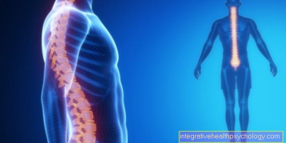
Scintigraphy of the bones
With the help of scintigraphy of the bones (also called skeletal scintigraphy), the bone metabolism can be visualized and areas with increased activity can be identified. Our bones are not lifeless scaffolds, but are subject to constant construction and dismantling. For the scintigraphy of the bones radioactively marked building blocks of the bone metabolism are used (diphosphonates). After the injection of the substance, it is distributed throughout the body and is built into the bones after a few minutes. The higher the metabolic activity, the more radioactive particles are incorporated and the more clearly a bone stands out in the image captured by the gamma camera.
This can be used for various questions that justify a skeletal scintigraphy. On the one hand, inflammatory processes and changes in the bones can be examined, such as in rheumatism or osteomalacia (softening of the bones). If there is a suspicion that a joint prosthesis has loosened, scintigraphy can provide information. If normal imaging (for example X-rays) does not provide any reliable information, it is still possible to examine whether a bone is broken or not. The question of whether the tumor has spread to the bone can also be investigated in patients with cancer.
However, the following must always be considered during the evaluation: The scintigraphy of the bones is very sensitive, which means that even small increases in metabolic activity can be reliably detected. On the other hand, the examination is not very specific, which means that no reliable statement can be made about the cause of an abnormality in the scintigram. A cancer patient who wants to examine whether malignant cells have spread into the bones can serve as an example. If the scintigram is normal, then a spread is also rather unlikely. However, if there are areas that are noticeable on scintigraphy, they do not necessarily have to be metastases (descendants of cancer). It can also be a more harmless cause, such as the result of a bruise. The assessment of the skeletal scintigraphy must therefore always be made individually in connection with other findings and circumstances of the patient. In addition to a scintigraphy of the entire skeleton, only part of the bones, for example the hands, can be examined in isolation.
Scintigraphy for rheumatism
In patients with rheumatic disease, scintigraphy can be used to examine the bones for inflammatory changes. In addition, this examination makes it possible to distinguish between pathological changes in the joints whether they are inflammatory or not. It is one of many possible examination methods for assessing the activity of the disease. However, scintigraphy is not suitable for diagnosing rheumatism because it is too unspecific.This means that although changes in the bones due to increased metabolic activity can be reliably detected, what is the cause cannot be determined by scintigraphy alone.
You might also be interested in: How do you recognize rheumatism?
Scintigraphy in children
A scintigraphy is also always a certain burden on the body radioactive material is located in the body and interacts there.
Therefore, scintigraphy is often avoided in children.
However, if there is a suspected case of Child abuse, scintigraphy can provide information here.
If a child is hit, it is usually not directly visible Broken bones.
But already that bruise of the bone and the surrounding tissue can be recognized with the help of the scintigram.
The reason for this is an increased Metabolic activity.
The struck area is supplied with more blood. Reason may be the bursting of a small artery be that too Skin bleeding leads.
However, a bruise is usually associated with increased blood flow.
The damaged tissue tries to regenerate and therefore needs more blood, which leads to increased blood flow and increased metabolic activity in the area of the bruise.
This increased activity can be detected in the scintigram.