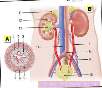
Figure ureter: A - cross section in relaxed state and B - retroperitoneal space with ureters (red)
- Ureter - Ureter
- Transitional epithelium - Urothelium
- Shift layer of the
Mucous membrane - Lamina propria - Inner longitudinal layer -
Stratum longitudinal internum - Outer longitudinal layer -
Stratum longitudinal externum - Middle ring layer -
Circular stratum - Connective tissue covering with
Blood vessels - Tunica adventitia - Aortic fork - Aortic bifurcation
- Rectum - Rectum
- Urinary bladder - Vesica urinaria
- Adrenal gland -
Suprarenal gland - Right kidney - Ren dexter
- Renal pelvis - Pelvis renalis
- Lower vena cava - Inferior vena cava
You can find an overview of all Dr-Gumpert images at: medical illustrations
$config[ads_text1] not found
Related images

Illustration
Ureteral stones

Illustration
Adrenal gland

Illustration
kidney

Illustration
Renal pelvis

Illustration
Pelvic inflammation

Illustration
Kidney stones

$config[ads_text2] not found
Illustration
Pain when
Urination

Illustration
Pain Abdominal Woman

Illustration
Pain Abdominal Man
Tags:
Orthopedics-Online Psychology Online Dermatology-Online Learning Problems-The- Drug