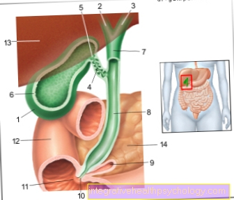
Figure gallbladder and large bile ducts, e.g. T. cut open, view from the front above
- Gallbladder Body -
Corpus vesicae biliaris - Right liver bile duct -
Ductus hepaticus dexter - Left liver bile duct -
Left hepatic duct - Gallbladder duct -
Cystic duct - Gallbladder Neck -
Collum vesicae biliaris - Mucous membrane -Tunica musoca
- Common
Liver bile duct -
Common hepatic duct - Main bile duct -
Common bile duct - Pancreatic duct -
Pancreatic duct - Extension of the united
Execution corridor -
Ampula hepatopancreatica - Large duodenal papilla -
Major duodenal papilla - Duodenum Descending Part -
Duodenum, descending part - Liver, diaphragmatic side -
Hepar, Facies diaphragmatica - Pancreas -
Pancreas
You can find an overview of all Dr-Gumpert images at: medical illustrations
$config[ads_text1] not found
Related images

Illustration
pancreas

Illustration
Colon

Illustration
Small intestine
Illustration
liver

Illustration
Upper abdominal pain

Illustration
Digestive tract
Tags:
Schnheitschirurgie Psychiatry-Online Surgery Online Ent Anesthesia-Online