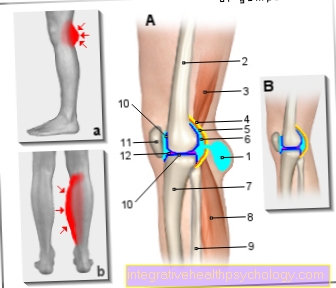
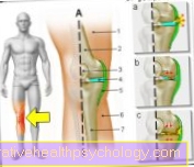
Illustration of the right knee joint from the right with Baker's cyst
- Baker's cyst
(Popliteal cyst) - Femur -
Femur - Semi-membranous muscle -
Semimembranosus muscle - Joint capsule, fiber layer
(yellow) -
Capsula articularis,
Membrana fibrosa - Joint capsule, soft layer
(orange) -
Capsula articularis,
Synovial membrane - Joint cavity
(filled with synovial fluid) -
Articular cavity, synovia - Shin - Tibia
- Internal calf muscle -
M. gastrocnemius, caput mediale - Fibula - Fibula
- Articular cartilage (dark blue) -
Cartilago articularis - Kneecap - patella
- Inner meniscus -
Meniscus medialis
A - Knee joint effusion with Baker's cyst
B - Healthy knee joint
a - swelling in the hollow of the knee
b - swelling in the calf muscles
You can find an overview of all Dr-Gumpert images at: medical illustrations
$config[ads_text1] not found
Related images
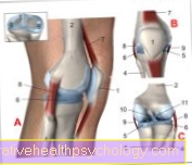
Illustration
Knee joint
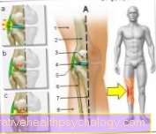
Illustration
Back of the knee pain
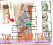
Illustration
Outside knee pain
Illustration
Inside knee pain
Illustration
Pain in the legs
Illustration
Pain in calf
Tags:
Psychology Online Internal Medicine Dermatology-Online Anatomy-Lexicon Pediatrics