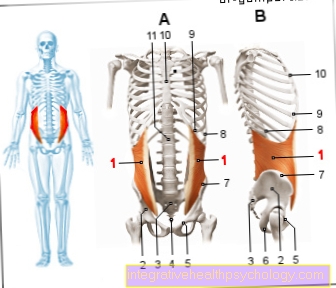
Illustration of the internal oblique muscle: chest from the front (A) and from the side (B)
Internal oblique muscle
- Internal oblique abdominal muscle -
Obliquus muscle
internus abdominis - Iliac scoop -
Ala ossis ilii - Sacrum -
Sacrum - Tailbone -
Os coccygis - Pubic bone -
Pubis - Ischium -
Os ischii - Iliac crest -
Iliac crest - 9th rib - Costa IX
- Costal cartilage -
Cartilago costalis - Sternum - sternum
- First lumbar vertebra -
- Vertebra lumbalis I
You can find an overview of all Dr-Gumpert images at: medical illustrations
$config[ads_text1] not found
Related images
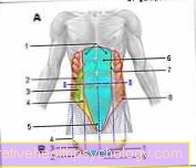
Fig. Outer oblique abdominal m.
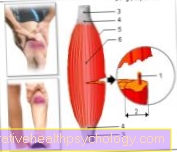
Figure muscle
Straight abdominal muscle
Illustration
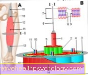
Muscle fiber
Illustration
Torn hamstring
Illustration
Muscles - abdomen
$config[ads_text2] not found
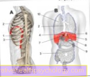
Illustration
diaphragm
Tags:
Ent Learning Problems-The- Schnheitschirurgie Neurology-Online Diagnosis