The ankle is made up of various partial joints.
The two largest joints are:
Together they form a functional unit and are called Articulatio cylindrica designated. The ankle joint is one of the most stressed joints in the body, as it has to support the entire body mass with every step.
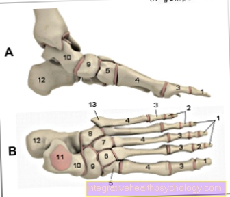
In addition to these, there are also smaller joints of the tarsal bones, which are, however, strongly fixed by ligaments and therefore hardly movable.


$config[ads_text1] not found
You can find an overview of all Dr-Gumpert images at: medical illustrations
$config[ads_text2] not found
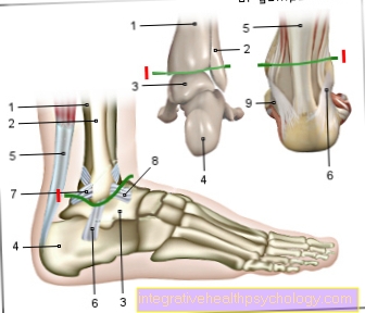
The upper ankle (Articulatio talocruralis) is made up of the articular surfaces of the Malleole fork and the Ankle bone (Talus) together.
The malleolar fork is inserted through the distal end of the Tibia (Tibia) - and Fibula (Fibula) educated. The ankle bone is enclosed by the malleolar fork from above on both sides and is therefore of crucial importance for the stability of the joint.
The upper ankle is a pure hinge joint and can with it just one movement To run. This consists in lifting the tip of the foot (Dorsiflexion) by approx. 20 ° and lower the tip of the foot (Plantar flexion) by approx. 30 °.
The Joint capsule surrounds the two ends of the Tibia and fibula, as well as that Ankle bone. This is where the Malleole fork (Outer and inner ankle) outside the joint capsule and are therefore very vulnerable to injury.
The joint in itself is further enhanced by various others Tapes fixed:

$config[ads_text3] not found
I - Upper ankle
(Joint line green) -
Articulatio talocruralis
You can find an overview of all Dr-Gumpert images at: medical illustrations
The lower ankle is part of the Foot and consists of:
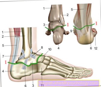
The boundary between these parts is formed by the ankle-calcaneus ligament (Talocalcaneum interosseous ligament). Both parts each have their own joint cavity, but from a functional point of view, the parts cannot be separated.
The anterior joint part consists of the interaction between the talus (Talus) and parts of the calcaneus (Calcaneus), Scaphoid (Navicular bone) and the plantar calcaneonavicular ligament (joint socket).
The rear part (Articulatio subtalaris) is due to the outwardly shaped side (convex surface) of the calcaneus (Calcaneus) and the inwardly shaped side (concave face) of the talus (Talus) shaped.
The lower ankle can cause the inner (Supination) and outer (Pronation) Lift the edge of the foot. Other joints are automatically moved with this movement, so that the entire pronation and supination movement of the foot is greater than the pure movement in the lower ankle joint.
The Range of motion for the combined movement is around 50-60 ° for the supination and around 30 ° for the Pronation.
The lower ankle is also affected by different Tapes stabilized:

I - Lower ankle
(Joint line green) -
Articulatio talocalcaneonavicularis
You can find an overview of all Dr-Gumpert images at: medical illustrations

Who am I?
My name is dr. Nicolas Gumpert. I am a specialist in orthopedics and the founder of .
Various television programs and print media report regularly about my work. On HR television you can see me live every 6 weeks on "Hallo Hessen".
But now enough is indicated ;-)
Athletes (joggers, soccer players, etc.) are particularly often affected by diseases of the foot. In some cases, the cause of the foot discomfort cannot be identified at first.
Therefore, the treatment of the foot (e.g. Achilles tendonitis, heel spurs, etc.) requires a lot of experience.
I focus on a wide variety of foot diseases.
The aim of every treatment is treatment without an operation with a complete restoration of performance.
Which therapy achieves the best results in the long term can only be determined after looking at all of the information (Examination, X-ray, ultrasound, MRI, etc.) be assessed.
You can find me in:
Directly to the online appointment arrangement
Unfortunately, it is currently only possible to make an appointment with private health insurers. I hope for your understanding!
You can find more information about me at Dr. Nicolas Gumpert
The ligament structures of the foot are particularly often affected by injuries. With a typical twisting of the foot inwards or outwards, damage to the capsular ligament apparatus with tearing, stretching or straining of the affected ligaments can occur.
Bony injuries, such as fractures of the outer or inner ankle, are possible, but rarely.
With around 20% of all sports injuries, the ankle is very often affected by trauma of all kinds. Compared to other joints, however, there are hardly any signs of wear and tear in the ankle, provided that no trauma has preceded it.
Thus, the most common osteoarthritis occurs after ankle sprained fractures or complex injuries to the capsule and ligaments.
The Joint between the calcaneus and cubic bone (Articulatio calcaneocuboidea) is a Amphiarthrosis, a very strongly fixed joint in which hardly any movements possible are. This joint is also fixed by additional tight ligaments.
Also the Tarsal-metatarsal joints (Articulationes tarsometatarsales) and the Metatarsal joints (Articulationes intermetatarsales) are Amphiarthroses and therefore hardly movable.
It will Metatarsophalangeal joint (Articulationes metatarsophalangae) and Toe middle or Toe joints (Articulationes interphalangae) differentiated.
The Basal joints are Ball joints, however, they are strongly fixed by various straps and are therefore hardly movable.
The Middle and end joints are Hinge joints and a little more agile.
The ankle is one functional unit consisting of:
The ankle is strongly fixed by ligaments and therefore does not allow many different movements.
Due to the high stress of the ankle, it must be very stable, this is ensured by the ligament and capsular apparatus.
However, since the outer and inner ankles are not located within the joint capsule, they are very prone to injury. This is how most sports injuries occur in the area of the outer or inner ligaments Ankle trauma. This can cause tearing, straining, or stretching of the ligaments. Despite the high stress on the ankle, signs of wear and tear (osteoarthritis) are very rare without a previous serious injury.