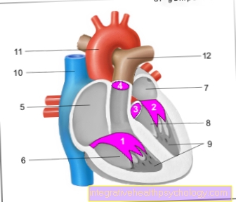
Synonym: Valvae cordis
The heart consists of four cavities, which are separated from each other and from the respective executing blood vessels by a total of four heart valves. This enables blood to flow in only one direction and only if it occurs as part of the heart's action (Systole or diastole) makes sense.
One differentiates the heart valves in two leaflet valves from two pocket valves.
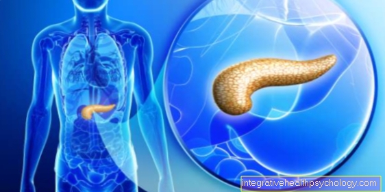
You can find an overview of all Dr-Gumpert images at: medical illustrations
$config[ads_text1] not found
The heart valves are in the so-called Heart skeleton, a fiberboard between Forecourt and chamber, anchored. They are protuberances of the Endocards, i.e. the innermost layer of the heart wall and ensure that the Blood in one direction only (unidirectional) by the heart can flow. Besides, they let you Blood flow only at certain times the heart action too. they also Function heart.
One differentiates between two Sail flaps (Valvae cuspidales) and two Pocket flaps (Valvae semilunares). The Sail flaps are also called Atrioventricular valves (AV valves) because they are between atrium (Atrium) and chamber (Ventricle) are located. The naming of the heart valves is based on the respective number of leaflets.
$config[ads_text2] not foundThe AV valves preventthat during the systole, in which the ventricle tenses, Blood back from the chamber to the atrium flows. The sail flaps are over Tendon threads (Chordae tendineae) with the Papillary muscles connected. These are anchored in the wall of the heart chamber and ensure that the valves do not recoil too far into the atrium when they close and during the tension phase.
The two Pocket flaps or Semilunar valves are respectively between the ventricle and the evacuating vessel.
Consequently prevent the pocket flaps den Return of blood from the two large vessels into the chambers after the systole has ended.
Their Names they have therefore that they are out 3 crescent-shaped each (semilunaris - crescent-shaped) Bulges or pockets.
$config[ads_text3] not found

The heart action can be found in diastole (Relaxation and filling phase) and Systole (Tension and expulsion phase) subdivide.
It was previously believed that the closure of the AV valves at the onset of systole was the first of the two Heart sounds would generate. However, it is now accepted that the 1. Heart sound only after the end of the AV valves, namely through the Tension of the ventricular muscles comes about.
The 2. Heart sound however, is actually a Key closing tone. It arises from the End of the pocket flaps at the end of the systole, i.e. after the blood flows out of the ventricles into the Lungs- or body circulation has been ejected.
Is the Function of a heart valve restrictedso this is called Heart valve vitium designated.
Such a Vitium can congenital or acquired be. There are two types of functional restrictions:
Slight valve defects can unnoticed stay, higher grade become usually symptomatic sooner or later.
Common to all valve defects is then the Exertional dyspnea (Difficulty breathing sometimes even with little physical exertion).
Most commonly affected are the Valves of the left heart, so the Mitral valve and the Aortic valve.
Read more about the topic here: Valvular heart disease