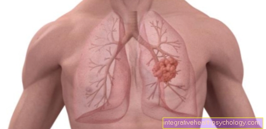
The medial meniscus is a Part of the knee joint. He has - in common with that External meniscus and the Cruciate and collateral ligaments - an important part of the smooth functioning of the knee movement.
In the case of an inner meniscus tear, it comes off accordingly Pain to a Function restriction the knee movement.

Meniscus lesions arise at younger people mostly through Violence on the knee, either as a result of one Trauma (e.g. in an accident) or, for example, by a jerky movement in sports, whereby the rotation of the knee with the foot fixed is the most common accident mechanism.
Hence come Medial meniscus tears especially common in sports like Soccer or Downhill skiing in front.
Since the Medial meniscus with the inner sideband of the knee joint grown together it has less opportunity to "evade" than the outer meniscus, so that it is essential in the event of an accident more often affected is than this.
A typical combination of injuries in the knee joint is the so-called "Unhappy triad" consisting of:
$config[ads_text1] not found
In humans from about 40 years Meniscus damage is common degenerative Nature, which means that in old age a natural wear and tear leads to the menisci already tear if there is little external force or even without it.
A distinction is made between different types of meniscus tears, each of which describes the shape of the tear:
Of these, the first two are the most common. In the case of a flap tear and basket handle tear, the part of the meniscus be pinched.
At a acute traumatic medial meniscus tear it becomes apparent immediately after the triggering event Pain in the affected joint space, besides is the Mobility immediately severely restricted.
At Entrapment of a portion of the meniscus can it become a Joint blockage come that knee can no longer be fully flexed or stretched. A effusion forms - if at all - slowly off because the Menisci poorly supplied with blood are and so not much with a tear blood emerges into the joint space.

Who am I?
My name is dr. Nicolas Gumpert. I am a specialist in orthopedics and the founder of .
Various television programs and print media report regularly about my work. On HR television you can see me every 6 weeks live on "Hallo Hessen".
But now enough is indicated ;-)
The knee joint is one of the joints with the greatest stress.
Therefore, the treatment of the knee joint (e.g. meniscus tear, cartilage damage, cruciate ligament damage, runner's knee, etc.) requires a lot of experience.
I treat a wide variety of knee diseases in a conservative way.
The aim of any treatment is treatment without surgery.
Which therapy achieves the best results in the long term can only be determined after looking at all of the information (Examination, X-ray, ultrasound, MRI, etc.) be assessed.
You can find me in:
Directly to the online appointment arrangement
Unfortunately, it is currently only possible to make an appointment with private health insurers. I hope for your understanding!
Further information about myself can be found at Dr. Nicolas Gumpert
$config[ads_text3] not found

It expresses itself very classically Medial meniscus tear by Pain. The pain intensity can vary and through Palpation, so through Feeling certain places and applying pressure, be reinforced. In meniscus lesions there is usually a Pressure pain at the respective joint space in front. This means that the pain in the case of a tear in the medial meniscus occurs in the Inside of the knee joint, so on medial joint space, are localized. Certain movements can also cause an internal meniscus tear "Snap or crack" noises occur. The reason for this is one Joint blockagein which the torn or torn part of the medial meniscus becomes trapped in the joint.
$config[ads_text2] not foundThis goes hand in hand with the Restriction of movement and decrease in resilience. Especially the Extension movement, that is, the stretching inhibited and particularly painful. But also Rotational movements can increase pain intensity; the pain of an medial meniscus lesion is through External rotation provoked.
All of this can be done regardless of the check special testsby moving the knee joint, i.e. alternately extending, flexing and rotating it in or out. The nature of the pain depends on whether it is a acute and fresh medial meniscus lesion acts or whether the Injury for a long time consists.
Sudden and severe pain speak for a acute happening with more severe lesions like a tear. However, if there has been an injury to the medial meniscus in the past without it being recognized and carried over, the can Pain worsens over time and with increasing damage occur.
$config[ads_text4] not found
Patients with a medial meniscus tear also report feeling the Instability in the knee joint. A common symptom of traumatic injuries is that swellingwhich, however, is not always typical of an inner meniscus lesion. Due to the loss of the shock-absorbing function, the knee joint surface is extremely irritated, which can cause a inflammatory process and a joint effusion with swelling as a result.
With regard to the traumatic causes based on sporting activity, strong shear forces, twisting or dislocating the knee, falls and an abrupt stop in movement lead to a meniscus tear, which is particularly common in sport. So it can be said that the sporting aspect plays an important role in relation to the causes of an inner meniscus tear.
The movements described, which can lead to a crack, occur mainly in sports such as tennis, squash, basketball, soccer and skiing. Jumps from great heights also put a knee joint under great stress and can provoke an inner meniscus tear.
Apart from acute traumatic causes, meniscus damage can also be incurred in everyday life. A typical dangerous movement is, for example, "crouching down".
In addition to the two main causes, the degenerative processes and traumatic events, a genetic, congenital variant of the meniscus also plays a role in the risk of an inner meniscus tear. The so-called "disc meniscus" is a clinical picture in which the menisci have a changed shape. Instead of the usual crescent-shaped appearance, the menisci, as the name suggests, rather have the shape of a disk. Certain pathological processes increase the risk of a lesion of the medial meniscus, especially during exercise and, in particular, of a medial meniscus tear.
After a medial meniscus tear, the affected joint space is clear pressure painful. To check whether it is actually a Medial meniscus tear is there several diagnostic meniscus tests:
The distinction between one Medial meniscus tear- and an external meniscus tear then happens with the help of the precise pain localization, whereby a increased pain in the internal joint space rather for one Tear of the medial meniscus speaks.
A X-ray image usually takes place on Exclusion of bone damage (e.g. in the form of a hernia, i.e. a fracture) Medial meniscus tear but cannot be seen in the X-ray image, this would be one MRI image (Magnetic resonance tomography or synonymous Magnetic resonance imaging) necessary.
Even with an MRI in the event of a meniscus tear, it cannot be diagnosed with 100% certainty.
Especially the small and degenerative cracks, so-called age cracks, are sometimes difficult to see in the MRI.
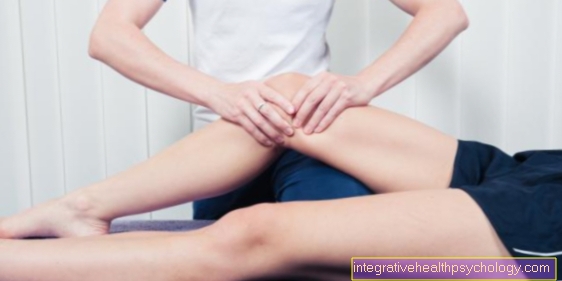
The patient lies on his stomach with one knee bent to 90 °. The examiner now fixes the patient's thigh with one hand or the leg. At the same time, he now rotates the patient's leg with the other hand, once under pressure and once under tension. It comes with the External rotation to pain, lies a Medial meniscus damage in front.
$config[ads_text1] not foundThis test is also called a Abduction and adduction stress test called. The examiner stabilizes the thigh of the patient lying on his back with one hand and grips the ankle region with the other hand. To test the medial meniscus, the upper hand grips the inside of the thigh or knee and the lower hand grips the outside ankle. Now the examiner bends and extends the leg while adducting it at the same time, i.e. under Varus stress puts. Of the Compression pressure the examination can trigger pain in the corresponding meniscus, in this case in the medial meniscus, and indicates its Lesion down.
For this test, the patient must be in a cross-legged position on the examination table. The examiner now pushes the knees, which are currently in an externally rotated and bent position, moderately strongly towards the surface. If the exertion of pressure in the medial joint space is felt to be painful, the patient probably has one Internal disc lesion in the posterior area.
The Steinmann I test aims at one Trigger rotational pain. The patient lies on his back with the knee flexed approximately 30 °. The examiner grips the lower leg with one hand and the heel with the other, from where he is once an internal rotation and then an external rotation performs. External rotation pain for one Damage to the medial meniscus.
In the second Steinmann test, the patient also lies on his back. The examiner tries to unite by feeling the medial and lateral joint space Trigger pain of the respective meniscus (medial pain = medial meniscus lesion). It is important that the Wandering pain point can: When flexing, the pain in the affected joint space moves backwards and when stretching it moves forward. In addition, the examiner rotates the patient's leg inwards and outwards while simultaneously doing one axial compression (i.e. pressure from below against the knee joint). External rotation pain speak here for Medial meniscus lesions.
Other functional tests that are not explained in detail are the Bragard test , of the Cabot test, the Childress-Sign of McMurray test (Fouche sign), of the Anderson compression test, of the Remember test, of the "Thessaly test", the Turner sign, of the Rotational compression test according to Pässler and the Tschaklin sign.
At chronic meniscus lesions can also be a Arthroscopy be indexed.

Meniscal tear
(= Meniscus rupture)
I - longitudinal tear
II - oblique view (rag tear)
III - radial crack (transverse crack)
IV - basket handle tear (special form)
V - degeneration (wear and tear)
You can find an overview of all Dr-Gumpert images at: medical illustrations

You can find an overview of all Dr-Gumpert images at: medical illustrations
That is far more common acute / fresh medial meniscus tear. That is true MRI (Magnetic resonance imaging) as the means of choice of imaging techniques for a medial meniscus tear. With the help of the MRI (or Magnetic resonance imaging) an inner meniscus tear can be assessed more precisely in terms of its shape and extent.
The principle of MRI is based on the magnetic property of individual atomic nuclei in our bodyeach a specific characteristic angular momentum to have. The exact function is very complex - simply put, a computer can record and evaluate the impulses so that in the end a three-dimensional image arises. The MRT is a 3D recording process, which Show meniscus tears in every spatial plane can. Through the use of magnetic fields and radio waves, the MRI image offers the possibility of different things Image contrasts. The MRI image is weighted depending on which tissue can be assessed.
Even if that MRI is the gold standard in the diagnosis of a meniscus tear is true CT (Computed Tomography) as a possible alternative. This is a X-ray procedure (ionizing radiation), which in comparison to the pure X-ray additionally Represent soft tissue can.
To a Fracture of the bony structures as a concomitant injury to a meniscus lesion to be able to exclude that conventional x-ray image in 2 planes on.
Read more on this topic at: MRI for a tear in the medial meniscus
There are two different approaches to treating an inner meniscus tear:
Both surgical methods of the medial meniscus tear usually result in a Extensive freedom from symptoms after a few weeks reached.
This is followed by immobilization for a few days of the knee joint before having a further physiotherapeutic treatment is started.
In addition to the previously mentioned therapies, taping can also be a sensible therapeutic approach for an inner meniscus tear. In the meantime, taping has become an established treatment method for an inner meniscus tear, especially for sports injuries, because it fulfills the task of a functional bandage.
The different colors of the tapes imply the strength, so that depending on the degree of severity, you can choose which tape is the right one. The tape supports the knee joint with its stabilizing function without restricting stretching or bending movements. The tape is very elastic and is very comfortable to wear. In addition, it protects against swelling in the knee joint area through slight compression. In addition, an improved blood circulation due to the slightly massaging effect of the tape on the skin should be noted.
The taping can be used as a pure conservative treatment as well as post-operative care be used. The choice of indication depends on the type and intensity of the symptoms. In the case of a large, total medial meniscus tear, taping is not sufficient to bring about complete healing of the medial meniscus. However, after surgery for such a severe meniscus tear, tape dressings can cause the Influence regeneration and the healing process through relief. In the case of a medial meniscus tear, conservative tape therapy can also be used primarily for Pain relief contribute. The reason for this is the fact that the knee joint is relieved by the stabilizing component and thus the damaged meniscus is also exposed to less pressure and friction forces.
In order to have a good therapeutic effect, it is very important that Apply tape correctly. The tapes should be placed in a bent position (approx. 70 °). In most cases, two strips are applied to the skin; one strip along the inside of the kneecap and the other along the outside. The kneecap should be free and not covered by the tape. You should inquire about the exact system technology from the attending physician.
Read more on this topic at: Tap your knees
In most cases, the damage is on Medial meniscus but so pronounced that one Conservative treatment is insufficient and therefore an operation is indicated. The goal of a medial meniscus tear is Preservation of the meniscus.
The operation is one arthroscopic surgery using an endoscope, which is made through small incisions in the Knee joint is introduced. You can use more small cuts Trocars with different surgical instruments be used.
Depending on the type and severity of the medial meniscus tear, it can be surgically treated in different ways: Basket handle crack or a crack at the base before, the surgeon sets one Meniscus suture. With the help of a special suture technique, the torn inner meniscus can be fixed again and correctly positioned so that it grows back together.
In addition to the meniscus suture, there is also the Meniscus resection and Meniscus transplant. The former, i.e. a resection or removal, is necessary if the Inner meniscus lesion too large is to be reattached using the suture. A suture may also not be possible if the tear is in an area that has poor blood circulation, for example. A however, good blood circulation is a prerequisite for good wound healing. Therefore a Partial removal of that meniscus region which may not be adequately supplied with blood.
A distinction is made between meniscus removal Partial and total resection. By definition, one removes a Partial resection of less than half of the meniscus (<50%); in the Total resection, self-explanatory, of the entire meniscus. A Meniscus transplant, So a Replacement of the menisci, is indicated when the patient's medial meniscus has already been removed. Especially young and still active patients benefit from this meniscus transplant as it reduces the risk of Osteoarthritis of the knee due to the insufficient shock absorber function can be lowered. In which Meniscus replacement it is either the Meniscus of a donor or around artificial tissue.