An MRT, i.e. a magnetic resonance tomography, is a high-resolution imaging method that delivers a three-dimensional image of the examined body areas without X-rays.
The patient is pushed into an elongated tube in which there is a strong magnetic field.
By stimulating the hydrogen nuclei in the body cells, the different components of the body are shown in a black and white picture.
With the help of an MRI of the temporomandibular joint, diseases and injuries in the area of the temporomandibular joint can be diagnosed.

The indication for an MRI scan is usually made very quickly and is often not absolutely necessary.
An MRI of the temporomandibular joint is often requested by an orthodontist if there is pain or problems in the jaw area.
Patients often complain of pain when moving the jaw, while chewing, or at rest.
Do you suffer from pain yourself after chewing?
$config[ads_text1] not found
In addition, misalignments of the jaw or a cracking of the jaw joint can occur when moving. The joint as well as the bones and soft tissues can be examined with the MRI. Congenital malpositions can thus be precisely differentiated.
In addition, an MRI can be performed to search for tumors, cysts or metastases.
With the help of the MRI image, the orthodontist can also better assess the possible access routes and risks during an operation.
Osteoarthritis is a degenerative disease in which the cartilage of the joint shows signs of wear and tear as a result of years of incorrect loading.
It usually occurs in older people, but a genetic variant can also occur in earlier years. Osteoarthritis usually manifests itself as pain in the area of the joint when chewing or moving - later also at rest.
Furthermore, the pain can also spread and lead to back and neck pain.
The diagnosis is usually made by an orthodontist using an X-ray.
Only in rare cases does it make sense to additionally perform an MRI of the temporomandibular joint.
There are several approaches to treatment.
An MRI examination of the temporomandibular joint begins with its preparation. First of all, the doctor informs you about the upcoming examination and informs you about the possible risks of an MRI examination.
It is not necessary to be sober before the examination.
In some cases, contrast medium is given beforehand via the vein to achieve better results. Since the MRI works with the help of a strong magnetic field, it is very important to remove all metal-containing parts on the body before entering the room. This also includes piercings, jewelry, cell phones, credit cards, etc.
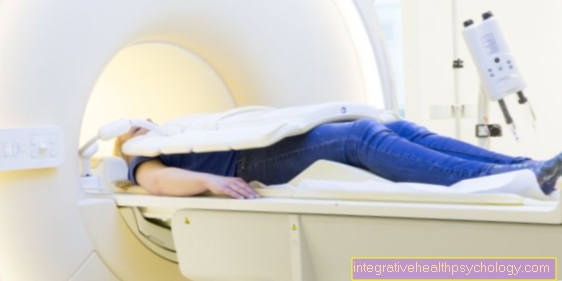
The MRI is an elongated tube with a hole in the middle through which a couch goes. For an MRI of the temporomandibular joint, the patient is driven headfirst into the tube until about half the torso is inside the tube.
When the MRI is on, it is usually very noisy, so patients always wear hearing protection and headphones. The examiner who is outside the room can also communicate with the patient via these headphones.
$config[ads_text3] not found
Since the patients are allowed to move as little as possible, they are often given a head frame with which the head can be precisely aligned and can no longer move. This is a big problem, especially for claustrophobic patients.
It may be possible to administer sedatives in advance.
Please also read our detailed articles:
$config[ads_text2] not foundAn MRI of the temporomandibular joint usually takes around 15 minutes.
However, the duration depends on the attitudes the examiner would like and on the cooperation of the patient.
In addition, there is the preparation time, i.e. the undressing of the patient, the positioning and the evaluation of the images.
A total of at least one hour should be planned for an MRI appointment.
In most cases, the evaluation is carried out by an experienced radiologist.
In some cases, experienced orthodontists can examine the MRI images themselves. The MRI provides a large number of sectional images in all 3 levels of the temporomandibular joint and the neighboring structures. These are displayed and evaluated on a computer.
A possible pathology can be precisely localized through the different levels. In the temporomandibular joint, the doctor assesses the joint surface, the bone substance of the upper and lower jaw and the neighboring structures.
Possible cysts or crooked teeth can also be diagnosed.
An MRI examination is usually a low-risk examination method. Since MRI works differently to an X-ray or a CT (computed tomography) without ionizing radiation, the body is not exposed to harmful X-rays.
This means that an MRI can also be used without hesitation in children or pregnant women.
$config[ads_text4] not found
In some cases, contrast agent is injected into a vein prior to the MRI. This can trigger allergies or intolerance, which can show up in the form of itching, rash, nausea or palpitations.
An MRI is a very narrow elongated tube. This is a big problem, especially for patients with claustrophobia, which is why the administration of a sedative is often indicated.
Since the MRI is a strong magnet, it is essential to remove everything that is magnetic beforehand.
If this is forgotten, the magnetic parts can fly through the room and injure the patient or damage the device.
The exact costs of an MRI of the temporomandibular joint cannot be precisely determined as they depend on various factors.
On the one hand, the patient's health insurance is decisive, i.e. whether they are privately or legally insured.
Other factors can also be included in the cost:
An MRI examination of the temporomandibular joint usually costs between € 400 and € 800.
Since an X-ray image is often sufficient to make a diagnosis, the patient has to pay for the costs himself.
In the case of an MRI of the temporomandibular joint, the administration of contrast agent is usually recommended, as the contrast agent can be used to better assess the soft tissue.
In particular, possible cysts or tumors in the area of the bone or the surrounding muscles can be better differentiated.
However, the administration of contrast media also involves risks, as allergies and kidney damage can occur.
During an MRI examination of the temporomandibular joint, the patient moves head first into the tube until the entire head is safely in the tube.
As a rule, the patient is therefore driven up to the upper body into the tube, so that half of the body is in the tube and the other half outside.
Claustrophobia or a defibrillator are contraindications to performing an MRI, so alternatives must be sought.
This depends on the question and the indication.
In most cases, an x-ray of the temporomandibular joint is always done first.
In most cases, this is also sufficient, as it allows the jaw joint and the bones to be displayed very well. However, the soft tissues cannot be examined properly with it.
Another alternative is CT (computer tomography).
This also works with ionizing radiation, but this allows the bone structures and soft tissues to be better represented.
It also provides sectional images and can help in finding a diagnosis.