X-ray examination, x-ray, x-ray, x-ray, x-ray
English: X-ray
An X-ray or X-ray examination is a method discovered by the physicist "Wilhelm Conrad Röntgen" in 1896 for x-raying the human body. With X-rays, the examination method is based on the different permeability of the tissue to X-rays.

Wilhelm Conrad roentgen accidentally discovered X-rays in 1896. This discovery still forms the basis of modern X-ray diagnostics and that developed from it Computed Tomography.
$config[ads_text1] not found
At the roentgen In the so-called X-ray tube, electromagnetic waves are generated by applying a voltage. These electromagnetic waves are also known as X-rays.
These X-rays are now aligned so that they leave the X-ray tube in the direction of the X-ray film. The classic X-ray film will be obsolete in the next few years and will be replaced by digital media (digital X-ray systems). However, the principle of operation remains exactly the same.
The object to be examined is now positioned between the X-ray tube and the X-ray film. X-rays are absorbed by tissue to different degrees. Bone tissue absorbs strongly, soft tissue less. As a result, the X-ray blackens to different degrees (X-rays blacken the image). So you have at roentgen a negative of reality.
At X-rays it is about electromagnetic rays, which are able to influence the matter they penetrate. The reason for this is the fact that X-rays ionizing properties exhibit. This means that they are able to Electrons (negatively charged particles) from atoms or molecules. As a result, positively charged particles are created.
$config[ads_text2] not foundIf X-rays hit human tissue during the X-ray, cells of the living organism can be permanently damaged. The X-rays emitted during the X-ray mainly affect the Genome of the cells hit. By releasing individual electrons, for example, the structure of the DNA contained base pairs changed. In most cases the organism is able to repair the damage caused by the X-rays through the action of the natural DNA repair system. With a correspondingly high radiation dose, however, such DNA changes can occur to such a large extent that a proper repair is no longer possible.
In Germany, the X-ray Ordinance and the Radiation Protection Ordinance regulate, among other things, the medical use of X-rays on humans.
According to this, an X-ray may only be performed if there is a so-called justifying indication (Healing display) was made.
This means that the health benefits of an X-ray must outweigh the damage from the radiation.
In view of the low radiation doses that are used in today's X-ray machines and the information content of fluoroscopy, this is almost always the case.
However, if equivalent methods with lower radiation exposure are available, these must be taken into account.
$config[ads_text3] not found
The justifying indication may only be given by experienced doctors with appropriate specialist knowledge if they can treat the patient personally on site.
However, non-specialist doctors may also prescribe an X-ray examination.
In this case, the performing radiologist assumes responsibility and can refuse the examination if he does not see the benefit of the treatment.
If a doctor provides a justifying indication without the necessary prerequisites, this can constitute bodily harm.
In a clinic, the radiation protection officer is liable for compliance with the law; in a sanatorium organized as an AG or GmbH, the managing director.
Failure to comply will result in fines. In practice, the problem arises that even inexperienced assistant doctors without the necessary specialist knowledge have to provide indications for an X-ray examination. This happens, for example, on weekends when there is no specialist in the house and, strictly speaking, violates the law.
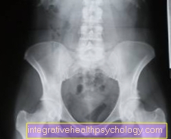
The native x-ray diagnosis, i.e. without the use of contrast media, is used primarily for questions relating to the skeleton.
Here it is the most informative method and involves comparatively little radiation exposure.
On the one hand, it is used to detect changes in the bones caused by injuries:
On the other hand, bone changes due to inflammatory processes (Inflammation of the bone marrow), Metabolic diseases (Misalignment of the fingers in gout), Tumors or degenerative diseases (arthrosis) can be recognized and monitored in their course.
$config[ads_text4] not found
In addition, the assessment of congenital malformations is an indication for an X-ray examination.
Another area of application for natural diagnostics is chest x-rays. The X-ray is a reliable diagnostic tool here because it shows the contrast between air and water well.
Read more on the topic: Chest x-ray (chest x-ray)
An indication is the external force on the chest: the X-ray provides information about broken ribs and cracks in the lung membrane, through which the lungs collapse.
Air, water retention and changes in tissue can be seen in the lungs. For example, an X-ray is indicated if pneumonia, tuberculosis, an increase in connective tissue, a vacuum or an effusion is suspected.
Heart diseases can also be identified and specified using an X-ray image: the extent of the individual heart spaces in the X-ray image allows conclusions to be drawn about the underlying disease.
In the abdominal area, the various organs differ little in their water content. As a result, the contrast of an X-ray is poor.
Other imaging techniques, e.g. Ultrasound, or tomographic methods, are superior.
However, there is an indication for the acute abdomen (life-threatening abdominal pain). Air or water retention and calcifications can be found.
An X-ray is also used diagnostically to detect stones in the urinary tract.
In mammography (X-ray of the breasts) you benefit from a very good resolution.
Certain details (Microcalcifications) can only be seen in X-rays.
An indication is therefore the suspicion (by touch or external changes) for tumor-like growth or the control of e.g. genetically predisposed risk groups.
If the X-ray is combined with the administration of contrast medium, it can also be used for other questions.
In the gastrointestinal tract, displacement of the organs, positional anomalies, and tumorous and inflammatory processes can be recognized and their course monitored.
The diagnosis of the small intestine is particularly important, as it is difficult to reach it with a camera.
The administration of contrast media is always associated with the risk of not inconsiderable complications and side effects.
As a result, the field of application of X-rays is being pushed back more and more by the new procedures - CT, MRT, ultrasound.
It is only displayed where there is (yet) there is no alternative or the question could not be conclusively clarified with other methods.
The catheter artheriography (The main artery is shown by inserting a catheter) for arterial occlusive disease, venography (Visualization of veins by injection of contrast medium) in the limbs if a thrombosis is suspected as well as the functional or structural examination of the urinary tract (by injecting or taking contrast media) if urine reflux, stress incontinence or obstructions are suspected.
An advantage of the X-ray compared to the cross-sectional imaging method is that images can also be taken during a movement (Esophagus when swallowing, ureter when urinatingn) can be made (dynamic x-ray examination or fluoroscopy).

The classic X-ray image:
There are different X-ray applications. By far the most common application is the classic one X-ray image.
The indication for use in the Orthopedics are questions that affect the bony support structure.
Many statements about the condition of bones and Joints do. The X-ray image is particularly helpful when it comes to questions about bone fractures and osteoarthritis of the joints.
However, the informative value of the X-ray examination is also limited. The cartilage can only be assessed indirectly. Soft tissue structures are generally not shown.
The fluoroscopy:
In addition to the classic X-ray image, there is fluoroscopy. X-ray fluoroscopy is particularly useful in orthopedics when the bone should be judged in its three-dimensionality. This is especially true in the surgery the case, e.g. when assessing fracture lines.
With fluoroscopy, less x-ray radiation is used and then projected onto a monitor via an amplifier so that the result is directly visible. In general, there is no permanent X-ray, but rather fast x-ray pulses are sent out. This allows the amounts of radiation to be reduced.
A fluoroscopy is more radiation-intensive than an X-ray image, depending on the fluoroscopy time.
Computed Tomography:
The Computed tomography (CT) is a special examination that developed from the X-ray examination. We have dedicated a separate chapter to this topic.
Contrast media:
X-ray contrast media are liquids that cannot be penetrated by X-rays. The result is that the image contrast increases. Contrast media are used in special issues disc prolapse, Intervertebral disc diseases and often used in the search for bone tumors partly in combination with computed tomography.
Please also read our topics:
$config[ads_text2] not found

X-rays are so-called ionizing rays. Ionizing rays damage the genetic material (DNA).
Due to natural radiation, we are exposed to ionizing radiation every day. The harmfulness of X-rays essentially depends on the location of the X-rays used.
Hands and feet are comparatively insensitive to radiation, while images of internal organs are more radiation-intensive.
The risk and benefit of a diagnosis are carefully weighed up in each case.
Particularly in the case of an existing pregnancy, the indication for an X-ray examination must be carefully checked.
In summary, the radiation risk of x-ray examinations is usually overestimated. The low radiation exposure should be compared with the risk of an overlooked disease.
Also read our topic: X-ray examination of the child
X-rays in orthopedics are used, for example:
The procedure of an X-ray examination is generally known. You should remember to remove all metallic objects (jewelry) so as not to endanger the assessment of the X-ray image.
The X-ray represents an extremely important step in the diagnosis of many diseases. For this reason, this form of imaging has become an integral part of everyday medical practice. Nevertheless, the decision to take an X-ray should not be taken lightly and the respective indication should be carefully considered. In addition, particular care should be taken to ensure that no duplicate recordings be made. This problem exists mainly in the area of Dentistry.
The x-ray is generally a safe procedure and the radiation exposure is quite low compared to the natural radiation exposure from the environment. Still can be special frequent x-rays lead to side effects. This diagnostic method does not speak of direct side effects, but radiation exposure can influence individual body cells.
In most cases, however, the effects of the cellular changes are only just beginning after several years in appearance. For this reason every patient should have one X-ray pass and carry it with you when you visit a doctor. Must in this pass all recordings made are noted become. In this way, unnecessary radiation exposure through repeated x-rays can be prevented.
One of the most decisive side effects of X-rays is the effect on the human genome. When a patient is frequently exposed to X-rays, it occurs at the DNA level Mutations. In most cases, these mutations can be caused by the natural DNA repair system of the body are repaired and damaged sections are restored. However, it comes because of one too high radiation exposure to damage this repair system or if there are several mutations at similar locations, a correct or complete repair is no longer possible. As a result, it can develop years after the actual exposure Tumors come.
Above all, a special form of X-ray that Computed Tomography, is considered particularly dangerous in this regard. This fact can be explained by the fact that a single computed tomographic image releases a significantly higher radiation dose than the production of a normal X-ray image. In addition, X-ray methods require a so-called Contrast media is administered, care must be taken that the patient's medical history is fully recorded.
This is especially important because the most common contrast media have one high iodine content feature. When administering an iodine-containing contrast medium to a person with Hyperthyroidism (Hyperthyroidism) can be a side effect thyrotoxic crisis be provoked. It is one of the most common direct X-ray side effects. The thyrotoxic crisis should be considered potentially life-threatening secondary illness viewed and the affected patient immediately admitted to a clinic.

Even during pregnancy, after accidents or certain illnesses, it may be necessary to take X-rays.
In the course of pregnancy, however, the respective indication of the X-ray must be carefully considered. Any unnecessary imaging must be avoided for the benefit of the child growing in the womb. This also applies to the preparation of dental x-rays.
Learn more at: X-ray during pregnancy
To reduce the risk of having to have an X-ray during pregnancy, preventive measures can be taken if you wish to have children. A dental example of such preventive measures is the survey of a comprehensive dental status with the preparation of an X-ray overview before the pregnancy. In this way, dental treatments can be concluded early and the development of inflammatory processes within the oral cavity, which are usually difficult to treat without X-rays, can be prevented.
X-rays during pregnancy should therefore be avoided if possible. Nevertheless, women who have to take x-rays during pregnancy should note that the risk of actual harm to the growing child is rather low. The radiation exposure of most x-ray examinations is simply not high enough to negatively affect the development of the unborn child.For this reason there is nothing to be said against urgently needed recordings, for example after an accident. This is especially true for body parts that are very far from the uterus. These include above all the arms, legs and the chest. Other parts of the body, however, are at increased risk due to their proximity to the uterus during pregnancy. For this reason, pelvic x-rays during pregnancy, for example, should only be carried out if there is a significant risk to the health of the mother and / or child if it is not done. X-rays of the intestines, urinary tract, and torso should also be carefully considered during pregnancy.
In general, the attending physician should be informed about the existing pregnancy. Before carrying out an X-ray examination, the specialist staff is even obliged to explicitly ask women of childbearing age whether they are pregnant. Putting on a special lead apron to protect against scattering X-rays is also useful outside of pregnancy. Although the risk to the unborn child is relatively small, attention should be drawn to the possible side effects of x-ray examinations during pregnancy.
In general, the risk of x-rays is slightly higher, especially at the beginning of pregnancy. During the first few weeks after fertilization of the egg cell, proper implantation of the fetus can be prevented by high radiation exposure. In later stages of pregnancy, X-rays can influence the maturation of organs in the growing child. In rare cases, deformities and malformations of individual organs or entire organ systems occur. The further the pregnancy is at the time of the x-ray, the lower the risk of malformations.
Furthermore, some doctors suspect that there may be a connection between the X-ray during pregnancy and later cancer in the child. However, these theories have not yet been adequately proven.
In summary, it can be said that X-rays during pregnancy should only be performed under certain indications, but are nowhere near as dangerous as is often claimed. In many parts of the body, X-rays are even completely harmless during pregnancy when wearing a lead apron. The risk of exposure to radiation during pregnancy should always be weighed against the risk of not taking the images.