Lung cancer becomes coarse in two different types divided.
The distinction here takes place on the histological (cellular) level: there is small cell and non-small cell lung cancer (lung cancer).
The group of non-small cell tumors, for example, consists of 30% so-called squamous cell carcinomas, 30% adenocarcinomas and many other sub-forms.

Lung cancer is the number one deadly cancer in men. For women, bronchial carcinoma ranks second behind breast cancer.
The greatest risk factor is still smoking. Due to the gender-specific change in the group of smokers, more and more women are also affected by lung cancer.
$config[ads_text1] not found
The diagnosis of lung cancer is often complicated.
A malignant tumor disease is usually only discovered late, when clinical signs already appear. In many cases, the symptoms are unspecific, so they can indicate several diseases, but usually worsen considerably as the disease progresses.
So-called paraneoplastic syndromes can occur, particularly in the context of small-cell bronchial carcinomas.
These are comorbidities caused by the release of tumor toxins or hormone-like substances by the cancer. The symptoms are varied and can lead the treating doctor on the wrong track.
This delays the diagnosis and the chances of recovery gradually deteriorate.
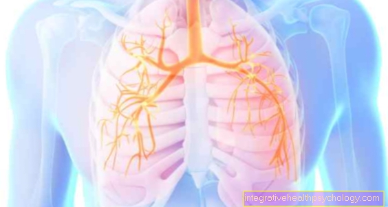
Imaging methods play the largest role in the diagnosis of lung cancer. In the X-ray image, the tumor foci can usually be seen as a shadow. However, the cancer must be big enough for that. In some cases, a tumor can be shown on X-rays before the first symptoms appear.
Computed tomography (CT) is the method of choice for further diagnostics. The CT determines the exact size and location of the tumor tissue. Otherwise, ultrasound and scintigraphy are used to search for metastases.
The laboratory values play a rather subordinate role in the detection of lung cancer. Since no conventional blood values are characteristically changed in bronchial carcinoma, so-called Tumor markers can be used. The markers to be examined are assigned to certain tumor types, but also occur in other types of cancer or diseases. The neuron-specific enolase (NSE) in small cell lung cancer, the carcinoembryonic antigen (CEA) in adenocarcinoma and the cytokeratin fragment 21-1 (CYFRA 21-1) in a squamous cell tumor.
At this point we would like to refer you to our main page on squamous cell carcinoma of the lung. You will find further important information on this at: Squamous cell carcinoma of the lungs
The Initial symptoms from Lung cancer are, if they occur at all, very unspecific.
to cough with or without ejection is a Signs of lung disease, however, a lung tumor is not primarily considered.
Pulls the Symptoms over a longer period of time, to step additional serious infections how Pneumonia on or will coughed blood several times, got to a malignant disease must be excluded.
The growth in size of an existing bronchial carcinoma can lead to pressure and occlusion in various organs. The bloodstream can be obstructed, it can become too difficulties swallowing come and Difficulty breathing during exercise occur.
Does the tumor infiltrate muscle or bone tissue, this is not just a sign of being advanced stagebut also associated with considerable pain.
The symptoms of a so-called paraneoplastic syndrome, which occurs more frequently in small-cell bronchial carcinomas, can be very diverse.
Through the hormone-like Substancesthat the tumor spreads, it can cause excessive production of other hormones, to Inflammation, to Fluctuations in the electrolyte balance, to Thrombosis and psychiatric disorders come with comprehensive clinical pictures.
$config[ads_text2] not found
$config[ads_text3] not foundTumors in the lungs above a certain size can be recognized by certain features on the X-ray. Tumors smaller than 1 cm are often overlooked, which makes early detection and a promising therapy more difficult.
Read more on the topic: Chest x-ray (chest x-ray)
The tumor tissue has a higher density than the lung tissue. The latter consists of air-filled alveoli and is dark in the X-ray. This means that few X-rays are absorbed, which makes sense in view of the less firm tissue.
The tumor tissue consists of many, densely packed cells. As a result, the tissue absorbs many X-rays and the tumor is lighter in the image than the surrounding tissue. Only X-rays that have passed through the body are recorded as the smallest dark point in the X-ray image. In the end, the interaction of the points results in the picture.
Tumors usually grow like a focal point in a round shape. If other lightened spots can be seen in the lungs, with their otherwise regular structure, in addition to the large bronchi, the examining doctor should be attentive. Malignant masses do not adhere to the anatomical limits of the lungs. They can cross the supply areas of the individual bronchi and even the individual lobes of the lungs. Some advanced lung tumors even grow beyond the borders of the organs and invade bone and muscle tissue or the pleura (Pleura) a.
The shape of benign and malignant tumors is significantly different.
While benign tumors have smooth edges, lung cancer grows infiltrating, i.e. penetrating. The cancer cells radiate into the surrounding tissue from a center. This is not displaced, as is the case with benign tumors or cysts, for example, but infiltrated.
$config[ads_text4] not found
Lime is often deposited in degenerate cell tissue. This shows up in the X-ray image as a radiopaque grain (white). So-called micro-calcification is a classic sign of a benign tumor, but it does not give the all-clear - even if calcifications very rarely occur in lung cancer, they are still possible. However, since calcium can be seen well in the X-ray image, this gives an indication of a change in the tissue. A targeted biopsy can now be performed, in which tissue is taken and then examined in the laboratory. X-ray diagnostics are the mainstay in diagnosing lung cancer and are often supported in the further course by CTs, as this is the only way to precisely determine the tumor boundaries.
in the Terminal stage from Lung cancer are the Symptoms usually already clearly pronounced. At least now you have Shortness of breath and Chest pain trained and increase the suffering of the patient massively. Due to the increased work of breathing and the mostly large tumor, much more energy is consumed than with a normal person The patient is losing weight extremely. Also fever and hoarseness can be symptoms of severe cancer.
As the bronchi are relocated, the lung sections that lie behind the tumor develop Inflammation, so-called retention pneumonia. she run away usually very difficult and do not respond well to conventional forms of therapy. The This further worsens the prognosis.