The function of the skin
introduction
The skin (Cutis) has two important functions for our body.
On the one hand it has a defense and protective function, on the other hand it is responsible for the absorption of stimuli. It has the function of warding off harmful effects and physiologically necessary exchange functions (Heat exchange) and to enable sensory impressions. The protective functions are very diverse.
1. Protection and defense functions
- Protection against mechanical effects
Is given to the skin by its tensile strength, elasticity and elasticity - Protection against chemical noxa and microbial intruders
The special structure of the granular layer and the skin surface film (e.g. fat content, pH 5.7, so-called protective acid mantle) form a barrier for the above. If pathogens or molecules should penetrate the skin, they trigger immunological defense mechanisms. - Protection against dehydration
The evaporation of water in a person without an epidermis would be 20 liters per day. - Protection against radiation noxa
The skin's radiation protection works by reflecting and absorbing light. - Immune reactions
Once noxae have overcome the skin's protective barrier, the skin can trigger an immune reaction. - Temperature regulation
The temperature is regulated reactively through blood circulation and perspiration. When it is hot, the blood vessels in the skin are expanded and water escapes to the surface of the skin.
2. The stimulus reception
Merkel cells are sensory receptors in the epidermis and are used as receptors for touch (Mechanoreceptors) viewed.
There are numerous nerves and nerve endings in the dermis.
Conduct free nerve endings
- pain
- Itching and
- temperature
A distinction is made between cold and heat receptors.
Father Paccini bodies are mechanoreceptors for pressure and vibration. They lie deep in the dermis or in the deep fat tissue of the palms and feet.
Meissner tactile bodies are touch receptors and are located in the connective tissue of the papillary layer of the dermis. They are also mainly on the palms of the hands and feet.
Depending on the localization of these corpuscles, they also have a different time behavior. Some with very fast to slow adaptation (Adaptation / habituation).
Illustration of the skin
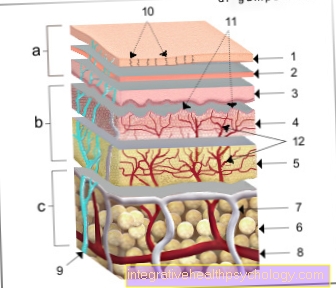
a - epidermis (1st - 3rd) - epidermis
b - dermis (4th - 5th) - Dermis
c - subcutaneous tissue (6.) - Tela subcutanea
- Horny layer - Stratum corneum
- Cornifying layer
(light layer
and granular layer) -
Stratum lucidum and
Stratum granulosum - Germ layer (prickly cell layer
and base layer) -
Stratum spinosum and
Stratum basale - Papillary layer -
Stratum papillary - Network layer - Stratum reticularre
- Subcutaneous tissue - Tela subcutanea
- Lymph vessel - Vas lymphaticum
- Artery - Artery
- Cutaneous nerve - Cutaneous nerve
- Duct of a sweat gland -
Ductus sudorifer - Papillae of the dermis -
Papillae (dermidis) - Vascular network of the dermis -
Subpapillary venous plexus
You can find an overview of all Dr-Gumpert images at: medical illustrations