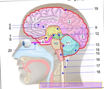
Gastroscopy
synonym
Gastroscopy
definition
A gastroscopy is a primarily diagnostic but also therapeutic procedure using an endoscopic camera to inspect the stomach and esophagus.

Indications
The gastroscopy is the technique of choice around Diseases of the To have the esophagus, stomach and duodenum checked. A gastroscopy can help to find the cause and the right therapy for the following complaints:
- Recurring heartburn
- Persistent nausea and Vomit
- Difficulty swallowing
- Chronic cough
- Pain in the upper abdomen
- Increased flatulence
- Unclear weight loss
- Vomiting blood
- Blood in the stool
- Gastric bleeding
In addition, if there is a suspicion of inflammation of the gastric mucosa, the gastroscopy can clarify the possible causes, such as an infection with Helicobacter pylori, an ulcer disease, a bulging or an injury to the mucous membrane.
In addition, doctors are able to provide a Injury or disease of the gastrointestinal mucosa available to treat them via gastroscopy. Bleeding in particular can be prevented by measures such as Placement of metal clips, rubber bands or injections of anti-bleeding drugs.
In most cases, a gastroscopy will rule out a Stomach ulcer performed because patients complain of chronic stomach pain before or after eating.
execution
Before a gastroscopy, the patient is prepared for this routine procedure.
This preparation includes one detailed explanation, during which the patient is explained the procedure and is informed about the risks and side effects. At the end of the explanation, the patient must give his consent and document this with a signature.
On the day of the gastroscopy, the patient must stay sober. Laxative measures are not necessary for this procedure.
If the patient is taking blood-thinning medication, consideration must be given to discontinuing them before the gastroscopy examination in order to avoid complications such as heavy bleeding.
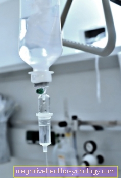
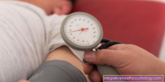
In addition, the patient receives a gastroscopy venous access placed in a vein, usually in the arm, on the day of the examination. This should be given fluids prior to the procedure in order to prevent the fasting patient from drying out (this is especially useful if the examination does not take place until noon).
A gastroscopy is usually done in one Short anesthesia with propofol carried out. Nowadays a small amount of the Anesthetic Propofol injected.
During the entire duration of the gastroscopy, the Oxygen saturation, breathing and Heart rate tracked via a monitor in order to be able to quickly initiate therapeutic steps in the event of a deterioration in vital signs.
During a gastroscopy, the patient is usually lying down in the left side position examined.
Before that, he is given a small rigid tube placed between his teeth. This tube is fastened behind the head with a rubber band. This becomes a Access to the oropharynx guaranteed regardless of whether the anesthetized patient clenched his teeth or not.
The examination instrument (Gastroscope) inserted into the mouth and throat.
The patient is not completely asleep and can also react to a limited extent to simple, loud commands.
The patient is asked to swallow as soon as the gastroscope reaches the level of the pharynx Larynx happens. If the patient swallows, the epiglottis closes the windpipe and opens the way into the esophagus, into which the gastroscope is now advanced.
At the tip of the examination device there is a very bright light, an opening through which air can be fed into the esophagus and stomach, and an opening through which samples from the tissue of the upper part are taken using small forceps and other instruments Digestive tract can be won. Furthermore, instruments can also be introduced through this opening, which can bring any bleeding to a standstill.
When inserting the gastroscope, air is first introduced into the esophagus in order to unfold the otherwise slack structure and to expose the view. The very strong light at the tip of the gastroscope allows an insight into the otherwise dark upper digestive tract.
First the gastroscope is maneuvered forward.
The actual inspection has not yet taken place here.
With a small regulator on the handle of the gastrocope, the tip of the device can be bent up to 180 degrees. This is the only way to guarantee that hidden areas can also be inspected. Different from one Colonoscopy the examination instrument can be pushed forward quite easily and only takes a few minutes.
As soon as the tip of the gastroscope has reached the stomach, the actual examination begins. The examination consists of three steps:
- inspection:
In all diagnostic gastroscopy, inspection is the most important part. Both the stomach and the esophagus are examined. Above all, the mucous membrane is examined and assessed whether it is reddened or inflamed, whether there are any sources of bleeding (both fresh bleeding, possibly spurting, and non-acute bleeding with older blood deposits) or whether there are unnatural constrictions in the esophagus and stomach. The stomach is also examined for gastric ulcers or abnormal tumors in the gastric mucosa.
When withdrawing the gastroscope, the esophagus is also viewed. In addition to bleeding, inflammation and redness, the so-called thrush (Fungal infection of the esophagus) and on varicose veins (Varices), which are very dangerous and can be an indication of a bypass cycle in the event of liver damage. - Biopsies:
From noticeable areas of the lining of the stomach small samples of skin taken in order to have them examined in the laboratory for a corresponding malignancy. For this purpose, small forceps are inserted from the outside over the gastroscope and pushed forward to the tip of the examination device. The forceps are placed on the suspicious area and the skin biopsy is removed and pulled outside. - Therapeutic approach:
In addition to the diagnosis of a gastroscopy, there is also the option to act therapeutically in the same session.
Especially in acute and spurting bleedingthat are seen in the esophagus or stomach, it is necessary to bring them to a standstill with the gastroscope. In most cases this works with one Clipwhich is brought in from the outside via the examination device and closes the bleeding vessel. Furthermore, the vessel can also be closed by an injection.
The gastroscopy procedure
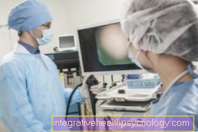
In most cases, the examination will take no more than a few minutes.
The process of a Gastroscopy is painless, but is often described as uncomfortable. The examination can be carried out while the patient is awake. Before starting, you can use a spray to clear the throat numb or at the request of the patient, tranquilizers (usually Midazolam or Diazepam) can be administered. These make the patient sleepy, so that he is not consciously aware of the process, but can sometimes still react to simple instructions. During the examination, the patient is positioned on the left side and a mouthpiece is inserted, which prevents the access to the throat area from being blocked by possible clenching of the teeth.
The examination instrument (Gastroscope) is an optical device that is made of plastic and is tubular. The guide is very flexible and has an opening at the end with a small camera and light source to gain good insight and to transfer the recorded images to a monitor. It also includes a channel through which instruments such as forceps or snares can be inserted during the examination and a channel through which air can be introduced. They are examined esophagus, of the stomach and the Duodenum. The endoscope gets slowly over the mouth of the patient towards throat introduced. When the examination device passes the throat, the examiner prompts the patient to swallow hard.
At the Swallowing process closes the Larynx the windpipe and thus prepares a clear path through the esophagus. Under visual control, the examiner pushes the tube downwards in small steps, past the lower esophageal sphincter, into the stomach. From there the hose continues over the so-called Stomach gatekeeper (Pylorus) in the duodenum (Duodenum) advanced. Once the deepest point has been reached, air is introduced through the endoscope to tighten the flaccid nature of the organs and thus a better view of the Mucous membrane to win. Then the endoscope is slowly returned to the patient. If you detect abnormal areas that show changes in the mucous membrane during the examination, you can take a tissue sample directly with a pair of pliers and then send it to the pathology department for further examination. In addition, possible bleeding can also occur during the examination through the endoscope by attaching a Metal clips or be breastfed by injecting medication.
Read a lot more information at: A gastroscopy procedure
Tissue sample
If the examiner recognizes during the Gastroscopy conspicuous areas in the mucous membrane, he can take a small sample of the tissue with the help of forceps inserted through the endoscope. The doctor grips the affected area with the forceps and snaps part of the mucous membrane with the tip of the forceps. The tissue can then be conveyed to the outside via the endoscope.
The sample is then sent to a specialized laboratory (pathology), where it is examined in the further course. The specialist makes small, thin layers of the sample delivered, which are colored with special coloring agents.
The nature and structure of the tissue is then assessed under the microscope. Particular attention is paid to the structure of the surface. You can see whether the mucous membrane is edematously swollen, whether inflammatory processes or bumps or defects can be seen. In addition, an assessment is made of whether there are any conspicuous cell aggregates that are differentiated from the rest of the tissue and that may indicate neoplasms, such as a tumorous change. The tissue sample sent in can be examined for any pathogens that may cause disease.
Read more about this topic here: biopsy
anesthesia
A gastroscopy can be complete without anesthesia done with easier Numbness, reassurance or under one brief anesthesia. Which variant is used depends entirely on the patient, his fears and his physical condition.
If the patient does not want to have anesthesia for a gastroscopy, the diagnosis is performed using the Throat numb. For this purpose, a spray is sprayed into the patient's throat and pharynx, which numbs the mucous membranes. This lets the patient hardly feel the tube or not at all suppresses the gag reflex.
There is also the option of having the patient in addition to the throat anesthesia Sedatives gets, for simple relaxation and resolution of fears (e.g. Midazolam or diazepam).
If the gastroscopy is to be performed under anesthesia, the patient first receives one peripheral venous access, preferably in a vein in the forearm. The anesthetic is usually supplied to the patient via this access Propofol, administered. During the entire examination, which the patient is under anesthesia, an employee monitors the Vital signs of the patient, i.e. his Pulse, Blood pressure, breathing, Oxygen saturation such as Cardiac activity about the EKG.
When performing a gastroscopy under anesthesia, it is important to remember that there are a few more general risks and that not everyone is suitable for anesthesia, e.g. owing to Allergies. Before the anesthesia, all factors should be discussed in detail with the anesthetist.
It must also be borne in mind that the Reactivity limited for some time after anesthesia and you are therefore not allowed to drive a car for 24 hours after an anesthetic in order not to endanger yourself or others.
Duration
The gastroscopy itself is a brief examination and usually afterwards 5-10 minutes past. The entire duration of the investigation is, however depending on the type of anesthesia. In the case of a gastroscopy under anesthesia, the preparation as well as the care after the examination require significantly more time. In this case, a time expenditure of about 2-3 hours to plan. It should also be borne in mind that after sedation, either with narcotics or in the form of anesthesia, it is much longer fatigue and Difficulty concentrating restrict in everyday life.
In the case of a gastroscopy without anesthetics, a Recovery time of at least 15-20 minutes given. If the patient is fine after this time, he can leave the practice / clinic afterwards. So all in all a Time expenditure of approx. 30 minutes.
Risks and Side Effects
Although gastroscopies are performed thousands of times a day and have long been considered a routine procedure, complications can still occur.
The most common side effect of gastroscopy are Flatulence after the procedure, as the air inflates the stomach.
It can rarely occur after a gastroscopy Intolerance reactions to the Anesthetics come. Here it is very helpful to inform the patient about appropriate before the procedure Allergiesthat have occurred in the past. In the event of an allergic reaction, the operation must be stopped immediately and the anesthesia withdrawn immediately.
Complications during and after the gastroscopy are also rare Bleeding that won't stop.
Minor bleeding On the other hand, they occur somewhat more frequently at the points where tissue has been removed with the biopsy forceps, but do not require any further action.
If there is excessive bleeding, a clip must also be placed at this point or the vessel must be injected under. In very rare cases, it becomes necessary to have open surgery to stop the bleeding.
In extreme cases it happens that the esophagus or parts of the stomach are pierced by the gastroscope (perforation). In this case, open surgery is almost always required to close the area again.
Complications
There are generally few risks associated with performing a gastroscopy, and it does occur hardly any complications on. Nevertheless, it is important to state the possible complications in advance of the examination. Since during the investigation of the Digestive tract When inflated with air, it can close immediately afterwards Flatulence come. Also a feeling of fullness and increased Eructation can occur. Mechanical irritation of the throat and larynx can also be temporary difficulties swallowing and hoarseness arise. Since the throat may be numb for a certain time after the examination, the patient should not eat food immediately after the examination. There is a risk that he will swallowed or food components get into the airways and there to one lung infection to lead.
Patients who have been given a sedative may have a depressant effect Circulatory problems arise. Especially with patients who have a anesthesia wish it can too allergic reactions come on the anesthetic. Therefore, allergies should be clarified in advance. If the patient has loose teeth, you may be able to Tooth damage arise. Both during and after the examination it is possible that bleeding as a result of Biopsies (Tissue samples) arise. In most cases, these are so small that they don't need any further treatment. However, major bleeding can also occur, which has to be stopped either with a metal clip or, in the worst case, with open surgery. Rarely, the wall of the digestive tract can also be pierced (perforation).
Cost of gastroscopy
The costs of a gastroscopy are usually covered by the health insurance company. In many cases, the procedure can be performed by a resident internist. Inpatient admission is sometimes necessary. Depending on this, the costs range from 100 to 400 EUR.
Gastroscopy in children
The gastroscopy also has one in childhood great diagnostic and therapeutic value. Be used for gastroscopy in the child very thin endoscopes used, which have a large resolving power. In contrast to gastroscopy in adults, the examination in children is in most cases carried out in anesthesia. During the preparation it makes sense to involve the parents, as they can calm the child and give him security. As a lot of children laying one Vein access To feel extremely uncomfortable and painful, it is possible to use the anesthetic in the form of a Anesthetic gaswhich can be inhaled.
The child is already asleep when the venous access is established. In addition to an age-appropriate endoscope and an appropriate anesthetic facility, an experienced doctor is required to carry out the examination. The procedure of the examination does not differ from the gastroscopy in an adult. In preparation, it is also important that no food has been ingested 12 hours beforehand so that there is no risk of the child choking on food components.
Read a lot more information under our topic: Gastroscopy in children
Summary
A gastroscopy is always performed when the patient described Discomfort in the stomach area or in the esophagus be seen.
After appropriate preparation of the patient, in which the person to be examined is informed about the risks and side effects, the patient is given a venous access through which an anesthetic is administered shortly before the procedure.
The patient must fast on the day of the examination.
The real investigation only takes a few minutes.
The patient receives the gastroscope, a tube with a camera attached to the tip and a light, through the mouth and throat and pushed through the esophagus into the stomach.
Air is passed through the gastroscope into the esophagus in order to expand it and to be able to see it better. The tip of the gastroscope can be moved by the examiner and thus enables a 180 degree view.
A gastroscopy consists of the inspection (examining the lining of the stomach and esophagus and looking for bleeding, tumors and gastric ulcers), Sampling (Biopsy of suspicious skin areas) and therapeutic options (If necessary, hemostasis using a clip and medication injections).
Even though a gastroscopy has become a routine procedure, it can still lead to complications Bleeding, perforations, infections, intolerance to the anesthetic agent exist and may require further surgical interventions.


.jpg)