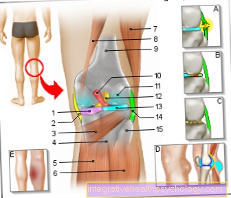
Illustration of the hollow of the knee: the hollow of the right knee with the knee joint and muscles

Back of the knee pain
A - tear of the outer band / inner band
B - injuries to the menisci
C - osteoarthritis
D - popliteal cyst / Baker's cyst
E - thrombosis

  1. Inner meniscus -
    Meniscus medialis
  2. Inner band -
    Ligament collateral tibial
  3. Popliteal muscle -
    Popliteus muscle
  4. Shin - Tibia
  5. Internal calf muscle -
    M. gastrocnemius, caput mediale
  6. External calf muscle -
    M. gastrocnemius, caput laterale
  7. Two-headed hamstrings
    Biceps femoris muscle
  8. Half-tendon muscle -
    Semitendinosus muscle
  9. Femur - Femur
  10. Posterior cruciate ligament -
    Ligamentum cruciatum posterius
  11. Articular cartilage -
    Cartilago articularis
  12. Anterior cruciate ligament -
    Ligament cruciatum anterius
  13. Outer meniscus -
    Lateral meniscus
  14. Outer band -
    Ligamentum collaterale fibulare
  15. Fibula - Fibula

You can find an overview of all Dr-Gumpert images at: medical illustrations

$config[ads_text1] not found

Related images

Illustration
Baker's cyst

Illustration
posterior cruciate ligament

Illustration
Torn knee ligament

Illustration
Inner knee ligament tear

Illustration
Knee joint

Illustration
Kneecap

Illustration
Outside knee pain

$config[ads_text2] not found

Illustration
Inside knee pain

Illustration
Cartilage damage

Illustration
Cruciate ligament

Illustration
meniscus

Illustration
anterior cruciate ligament


Tags: 
  • Orthopedics-Online 
  • Gynecology And Obstetrics- 
  • Dermatology-Online 
  • Psychiatry-Online 
  • Neurology-Online 
  • Prefer

    $config[ads_kvadrat] not found

    Preferences Categories

    $config[ads_kvadrat] not found

    Point Of View

    $config[ads_neboscreb] not found
    Top