Under a anterior knee joint pain refers to a pain that is mainly (not always exclusively) concentrated in the anterior part of the knee. This includes pain in the anterior thigh and lower leg, the kneecap, the quadriceps and patellar tendons and the anterior knee joint space.
Anterior knee joint pain can be caused by direct damage to the anatomical structures involved, or as transmitted pain in the case of damage to an anatomically distant location that is not causally a disease of the knee joint.

Below is an overview of the most common causes of pain in the front of the knee.
$config[ads_text1] not found
Quadriceps Tendon Disorders:
Causes of the kneecap (= patella):
Patellar tendon diseases:

Who am I?
My name is dr. Nicolas Gumpert. I am a specialist in orthopedics and the founder of .
Various television programs and print media report regularly about my work. On HR television you can see me every 6 weeks live on "Hallo Hessen".
But now enough is indicated ;-)
The knee joint is one of the joints with the greatest stress.
Therefore, the treatment of the knee joint (e.g. meniscus tear, cartilage damage, cruciate ligament damage, runner's knee, etc.) requires a lot of experience.
I treat a wide variety of knee diseases in a conservative way.
The aim of any treatment is treatment without surgery.
Which therapy achieves the best results in the long term can only be determined after looking at all of the information (Examination, X-ray, ultrasound, MRI, etc.) be assessed.
You can find me in:
Directly to the online appointment arrangement
Unfortunately, it is currently only possible to make an appointment with private health insurers. I hope for your understanding!
Further information about myself can be found at Dr. Nicolas Gumpert
A patellar dislocation can be congenital, habitual (habitual) or rarely traumatic due to twisting. In either case, the kneecap is not in the correct alignment with the rest of the knee. Mostly it is shifted sideways.
Only the traumatic luxation of the patella is associated with severe pain. The leg is kept in a flexed position and a strong joint effusion often forms. The cause of the dislocation is the damage to the extensor tendon apparatus located on the inside of the knee. Sometimes parts of the bone or cartilage can also be injured. In order to recognize this, an X-ray image and an MRI of the knee joint are made.
In the case of minor injuries, the doctor can reposition the kneecap by hand and then immobilize it in extension for a few weeks. In the case of larger injuries, the extensor tendon apparatus must be treated in a small operation (arthroscopy).
$config[ads_text3] not found
Sindig-Larsen disease (also Sindig-Larsen-Johansson disease) is a disease of the kneecap.
Overloading causes inflammation on the kneecap. This arises where the patellar tendon attaches to the kneecap. In the event of prolonged overload, the inflammatory reaction can cause severe damage to the patellar tendon (up to the tendon rupture) or to the kneecap. Small pieces of bone can loosen on the kneecap, which then die off because there is no longer any blood supply. So-called necrosis of these bone parts occurs.
$config[ads_text2] not foundThe main symptoms are pain in the anterior knee joint, usually just below the kneecap.
The quadriceps are our large muscle on the front of the thighs. As the name quadri (= four) - ceps (= head) suggests, it is a four-headed muscle, which therefore consists of four muscle parts. The quadriceps are the only muscle that causes extension in the knee joint.
The quadriceps tendon pulls at the top of the kneecap and is usually very thick and strong. Therefore, a quadriceps tendon tear is very rare. Such a crack can, however, arise if very high forces are applied (for example in high-speed accidents). A weakening of the tendon due to permanent excessive strain or inflammation in the knee joint can also promote a quadriceps tendon tear.
The result is pain in the front of the knee joint, usually above the kneecap. Usually, such a quadriceps tendon tear must be treated surgically.

$config[ads_text4] not found
Read more about kneecap arthrosis at:

You can find comprehensive information under our topic:

$config[ads_text1] not found

Read more on this topic:


$config[ads_text2] not found
Read more about this:
The patellar tendon is located below the kneecap. Therefore, pain immediately below the kneecap is often due to damage to the patellar tendon.
For example, excessive stress can lead to inflammation of the tendon, which is noticeable as anterior knee pain below the kneecap. People who work a lot on their knees (e.g. tilers and other craftsmen) are particularly affected. Diseases of the kneecap such as Sindig-Larsen's disease or the patellar tendon, e.g. Osgood-Schlatter's disease can trigger the symptoms.
The quadriceps tendon attaches at the top of the kneecap, so pain above the kneecap is often triggered by the tendon or the quadriceps muscle.
An injury to the muscle such as a torn muscle fiber or simply sore muscles or tension can lead to pain above the kneecap. Injuries to the tendon, such as a quadriceps tendon (torn), can also cause pain. Likewise, knee pain above the kneecap can be triggered by inflammation at the top of the kneecap. This is often caused by overuse and can even cause bone damage.
Swelling of the knee is a common symptom of pain.
On the one hand, swelling such as water retention in the knee itself can cause pain; on the other hand, the swelling can also be the expression of an injury in the knee joint. For example, if there is inflammation of the kneecap or neighboring tendons and ligaments, the inflammatory reaction causes swelling. A tear in a tendon, muscle fiber or ligament can also manifest itself as swelling and pain. Such a tear often leads to bleeding, which brings additional volume to the knee joint and therefore makes it swell.
Read more on this topic:
Water in the knee is a typical symptom of degenerative diseases. It often occurs with arthritic changes in the knee joint.
Injury to muscles or the menisci can also cause extra fluid in the knee. The destruction of the tissue causes the liquid to escape from the cells. This fluid remains in the knee joint and is colloquially referred to as water in the knee. The increased fluid accumulation often limits the mobility of the joint. In addition, pain can occur, especially when moving, as the water is displaced from the joint space and presses on other structures.
You might be interested in these topics:
The occasional pop in the knee occurs in many people and is usually not due to dire causes.
However, if the cracking occurs regularly with certain movements or if it is associated with pain, the cause should be clarified. A cracking can be triggered, for example, when the tendon of a muscle jumps back and forth between two positions when the knee joint is moved. But it can also be triggered by injured structures that block the knee in one movement, for example. If the joint is moved against this resistance, it will make a clicking sound.
Restriction of movement in the knee can have many causes.
Often there is pain when moving due to occlusion (e.g. osteoarthritis) or injuries to muscles, ligaments or menisci, which is why the knee can only be bent or stretched against pain. Water retention can also cause movement restrictions due to the increased volume in the joint. When a proper blockage occurs in the joint, an injured structure such as a torn meniscus is often the cause.
Loss of strength in the knee joint is usually due to muscular problems.
For example, overloading the muscles can lead to a temporary loss of strength. Injuries and inflammation also reduce muscle strength. Diseases of the nerves rarely lead to strength restrictions. Sometimes pain caused by other structural damage can also lead to reduced mobility and, over time, to a reduction in muscles and thus strength.
When climbing stairs, the knee joint is subject to particular stress.
The front part of the knee joint is particularly affected when walking down. Front knee pain when climbing stairs is therefore quite unspecific. They can indicate structural damage such as a tear in the meniscus or ligament and tendon disease. But arthritic changes and cartilage damage can also make themselves felt through front knee pain when climbing stairs.
If the anterior knee pain occurs after exercise, this usually indicates that the joint is overstressing.
If the pain occurred after a fall or collision, the knee should definitely be examined by an orthopedic surgeon for serious damage. If the complaints are more chronic, inflammation of the tendons or muscular overload can be the cause.
Therapy for pain in the knee joint is depending on the underlying cause. Is it a disease that conservative (non-surgical) can be treated, so help in the acute phase pain reliever drugs in tablet form (for example diclofenac, ibuprofen) or as an ointment (Voltaren, contains the active ingredient diclofenac). One often helps with injuries Cool of the knee while in other diseases such as osteoarthritis mostly warmth is perceived as more pleasant. Depending on the disease, a temporary full to partial Immobilization or support the knee joint in the form of a Orthosis to be necessary. The indications include, for example, injuries to the Tape apparatus or the menisci.
In the case of most illnesses or injuries, however, you should avoid sparing the knee for too long, so that it should be practiced again quickly - if necessary under physiotherapeutic guidance. Also Shoe insoles and Bandages can be helpful as well as Crutches.
In addition to physiotherapy, physical therapy is also used in conservative treatment. She works among other things with ultrasound as well Alternating and direct current. It comes to one improved blood circulation, one Inhibition of inflammatory processes and the Loosening of tense muscles. Invasive therapy methods for knee damage include Knee reflection (Knee arthroscopy) and surgery. Arthroscopy is often used for diagnostic purposes, but it also enables interventions such as one to be carried out Cartilage smoothing, or the removal of certain structures. The open surgery comes for example with a replacement of ligaments, a correction of Bone malpositions or using a artificial knee joint for use.
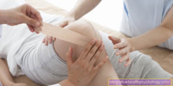
There are a number of different bandages for the knee joint that can be used depending on the type of discomfort or injury.
For the anterior knee pain, bandages that hold the kneecap in particular are often helpful. If only the kneecap is problematic, a patella bandage may also be sufficient. If the pain is muscular or caused by ligament structures, a larger bandage should be selected.
Further information on this:
Knee joint pain can have many causes. Accordingly, if symptoms persist, a doctor - usually an orthopedic surgeon - should be consulted to get to the bottom of the cause. At the beginning of the diagnosis there is always the questioning of the patient (anamnese). In the case of knee pain, important questions are, for example, where exactly it is located, when it first appeared, whether it is permanent or only in certain situations, whether it only occurs during exercise or also at rest, whether the pain radiates, whether it is in the There was a previous history of trauma, whether the patient is active in sports and, if so, which sports he does, what kind of occupation the patient does, whether he feels a sense of instability and what therapeutic attempts have been made so far, for example taking pain reliever medication.
Next up is the physical exam. The doctor should only inspect, i.e. look at the knee. Attention should be paid to malpositions, redness, knee swelling and bruise marks as well as the gait pattern. The investigation follows. It should be started carefully and if possible not directly at the most painful point, as this can severely limit the patient's willingness to cooperate.
When examining the knee, there are numerous tests called proper names that can differentiate more precisely where the problem lies. At the beginning, the range of motion should always be carried out according to the neutral-zero method in order to find out whether there is a movement restriction or not. More specific tests such as Steinmann 1, Steinmann 2, Apley and Payr can then be performed if damage to the meniscus or anterior and posterior drawer is suspected if damage to the cruciate ligament is suspected. After completing the anamnesis and the clinical examination, the examiner already has a suspicion in many cases and can adapt the further procedure to this suspicion.
Depending on the suspected cause, the following examinations can follow: a blood sample to find out whether it could be an inflammatory process, the recording of an X-ray image to assess the bony structures, a cross-sectional image examination (mostly magnetic resonance imaging = MRI of the knee joint) for an exact assessment of Soft tissues, ligaments and menisci in the knee joint area.
The minimally invasive diagnostic procedures include, in a broader sense, the knee joint puncture in the case of effusion and the arthroscopy, both of which can provide more precise information about the cause of the pain. During arthroscopy, in addition to inspecting the knee joint, interventions can also be made in the same procedure if necessary.
The diagnosis thus runs from the anamnesis and clinical examination to imaging procedures and minimally invasive interventions. For each patient, it must be weighed individually which examinations are sensible and which are avoidable. However, the history and clinical examination should be given to every patient without exception.
You might also be interested in: Chronic knee pain
Using our “Self” diagnostic agent is simple. Follow the respective link offered, where the location and description of the symptoms best match your symptoms. Pay attention to where on the knee joint the pain is greatest.
For the exact anatomical assignment, we refer to the Anatomy Lexicon our page.
Here are some key terms related to knee pain: