An MRI scan of the abdomen (also called an MRT abdomen) is one of the medical imaging techniques.
MRI is called magnetic resonance imaging or magnetic resonance imaging.
The abdomen is the medical term for the abdomen.
Depending on how many hydrogen atoms a certain body tissue contains, it becomes the final MRI image shown differently. So it is possible to use the different organs of the abdomen (abdomen). With the MRI all parts of the body can be examined without exposing the body to harmful radiation. The MRI especially for showing the soft tissues. To assess Bone structures is the CT (Computed tomography) more suitable.
$config[ads_text1] not found
The MRI examination of the abdomen denotes a representation of the abdominal cavity by means of MRI. It can be recommended and carried out by a doctor in various cases.
A MRI examination can be done by a doctor's prescription if various diseases are suspected.
Usually the MRI to diagnostic It is used for purposes, but it is also used to assess the progress of various diseases, for example to assess the effectiveness of cancer therapy.
Examples where a MRI examination of the abdomen include:
Means MRI of the abdomen can for example stomach-, liver-, Kidneys- and Pancreatic tumors or gynecological Tumors to be clarified.
$config[ads_text2] not foundIn some cases no MRI examination be performed. As a strong Magnetic field affects the patient, he may no metal parts wear on or in the body. Contraindications are therefore piercings, tattoos containing metal, metal implants, implanted defibrillators, Pacemaker and mechanical heart valves (with exceptions).
Read more on the subject here MRI and tattoos.
Is for the MRI examination Contrast media necessary, the Kidney function be checked. At insufficient kidney function may no contrast agent administration respectively.
Also in the pregnancy should be avoided.
If you are intolerant to iodine, you should not use a contrast agent containing iodine. As a rule, however, contrast media containing iodine are no longer used today.

If metal parts remain in or on the body during the examination, a Detaching these metal parts come through the magnetic field. This can lead to a Dislocation of implants come with injury to neighboring structures.
$config[ads_text3] not found
Free metal parts can be used in the MRI machine be attracted by the magnet and thereby to Injuries of the patient. Metallic make-up and tattoos can heat up due to the magnetic field and Tingling sensations up to Burns trigger.
It can also be Administration of contrast medium to an intolerance reaction, which in the worst case to a allergic shock can lead. There may be minor side effects after administration of contrast medium a headache, Skin tingling, malaise or feelings of warmth or cold. However, this is usually harmless and goes away after a short time.
$config[ads_text2] not foundPeople with Claustrophobia or excessive noise sensitivity may feel uncomfortable during the examination because the tube of the MRI is quite narrow and loud noises occur repeatedly during the examination. This should be discussed with a doctor in advance.
Please also read our topic: MRI for claustrophobia
Damage caused by the MRI examination per se are not to be expected, since there is no use of radioactive radiation. According to current knowledge, the MRI no harm to the unborn child is to be expected.
The private health insurance companies usually assume the cost of the MRI examination.
Both statutory health insurance companies is a corresponding indication required for the MRI is taken over. Otherwise the costs must be borne by the patient himself.
The costs are different in this case. Usually with 300 - 600 euros be expected.
You can find more detailed information on this under our topic: Cost of an MRI examination
$config[ads_text4] not foundWith a pictorial representation of the Gastrointestinal tract it is necessary that the patient come to the examination on an empty stomach. He is asked to do this the day before laxative to take in. From the evening before, only clear liquids be taken so that the Intestines remains free of stool residues and the intestinal structures in the MRI scans are visible.
On the day of the examination, it is important that the patient is informed before entering the Examination room all Discards metal parts. This also includes hair clips, piercings, earrings, all jewelry, keys and belts.
Carries the patient Metal parts in his body, for example Implants, this will be discussed in advance and it will be checked whether the MRI in this case can be done at all.
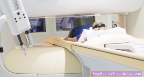
If these general precautions are taken, the patient can enter the examination room. The patient now lies down on the table of the MRI, which is moved into the magnetic resonance tomograph after correct positioning of the person to be examined.
If contrast medium is required for the examination, it is injected into the patient beforehand.
In the case of claustrophobia or general excitement, the patient can also be given a sedative.
To that Hearing to protect, the patient also receives headphone or ear plugbecause loud noises occur repeatedly during the examination.
The patient will then be out of the MRI driven out and can leave the examination room. Special precautionary measures are usually necessary after the examination.
The duration of the MRI examination may vary. It ranges from a few minutes to an examination duration of over an hour, depending on how large the area to be imaged is. Measurement pauses are inserted between the individual sequences, which extend the duration of the examination.
As a rule, an examination time of 30 minutes must be expected. During the duration of the examination, one should move as little as possible so as not to reduce the image quality.
In order to better display the desired structures in MRI to achieve, must in some cases Contrast media can be used.
This is usually done via a vein administered. In the MRI examination of Gastrointestinal tract however, it may become necessary to use contrast medium drink.
This then wets the directly Mucous membranes of Digestive tract and leads to a better representation there. So that the wetting of the Mucous membranes is as uniform as possible, the liquid should gradually within about an hour be taken.
It can take some time for the contrast agent to actually arrive everywhere. Hence the MRI examination do not take place immediately after drinking the contrast medium. Under certain circumstances, the high fluid intake with the contrast medium can subsequently result diarrhea occur. However, this is to be rated as harmless and disappears again within a short time.
$config[ads_text1] not found
Please also read our topic: Contrast media and MRI with contrast agent
The kidney can be assessed very well by means of an MRI examination of the abdomen. For example, changes in the organ structure, tumors or other anomalies in the kidneys and urinary tract can be recognized in the prepared sectional images. For this purpose, a contrast agent can also be administered that improves the image quality. With regard to the administration of contrast medium, the patient's kidney function must be taken into account, since the contrast medium cannot be excreted well if the kidney function is impaired. In addition, the kidney can be further damaged by the contrast agent. If the kidney function is intact, the MRI examination with contrast agent should not be classified as problematic.
Read more on the subject at: MRI of the kidney

In the MRI examination It is only necessary to be sober when examining the abdominal organs. Other MRIs usually do not require fasting.
Especially when recording the Intestines it is important that the patient is absolutely sober, otherwise the intestinal walls cannot be assessed. For this reason, one day in advance of such an examination laxative drank. From the evening before, only clear liquids be consumed so that the bowel remains free of stool remains. As part of the examination, the patient is then given a contrast medium and a liquid that unfolds the bowel. Sometimes an agent is also given that calms the intestinal motor skills so that the image quality can be increased. Following the examination, you can usually eat normally again.
The Sellink MRI examination is a special one MRI examination of Small intestine. Duodenum and Large intestine can easily be seen with the endoscope, the rest of the Small intestine however, is means endoscope unavailable, so that MRI must be used.
This is possible with the Sellink technology. It is mainly used for clarification inflammatory bowel disease applied, for example Crohn's disease or Ulcerative colitis. So that the Small intestine in the MRI can be easily assessed, laxative measures are necessary before the examination:
A light breakfast can be taken the day before, after which it must be removed so that the bowel is free of stool remains for the examination. On the day of the examination itself, the patient must sober stay. At the beginning of the examination, the patient receives a contrast medium, which over a to Small intestine advanced probe is applied. The contrast agent now wets the Mucous membrane and does this later MRI image visible. So that the intestinal walls do not lie against each other, one must now Methyl cellulose mixture administered through the tube. This expands the intestine so that it can be easily assessed in all its parts. Images are already being taken while the two solutions are being administered. You can also Intestinal peristalsis disorders be recognized.
Overall, the process is low-risk. The methyl cellulose solution can cause it in some cases Flatulence and stomach pain come, which, however, pass by themselves after a short time.
Further information can be found in our chapter: Sellink MRI examination
Also the stomach can by means of MRI be assessed. For example Tumors of Stomach or anatomical abnormalities be recognized.
For the specific investigation of the Gastrointestinal tract in the MRI however, special recordings are necessary in order to Mucous membrane to be able to judge better. The MRT examination according to Sellink can be used in which contrast agent is applied directly into the gastrointestinal tract. This will make the Mucous membrane directly wetted. The intestine is then stretched by a methyl cellulose mixture so that the intestinal walls do not lie against each other. Then the structures in MRI be assessed. To target the Gastric mucosa however, one is suitable for judging Gastroscopy (gastroscopy) better than one MRI examination.