The short neck muscles belong to the so-called autochthonous back muscles and are located to the right and left of the vertebral bodies of the spine. Their task is to maintain the vertebral bodies and to move the spine. The short muscles in the neck area are also stabilizing, but also make a significant contribution to the movements in the neck and head area:
Especially tilting your head forward and
this muscle group is used to put the head back (reclining).
The short neck muscles also play a key role in lateral head movements.
The short neck muscles include the rectus capitis posterior minor muscle, the rectus capitis posterior major muscle, the superior obliquus capitis muscle, and the inferior obliquus capitis muscle.
$config[ads_text1] not found
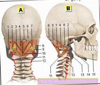
Illustration of the neck muscles
Figure neck muscles:
A - rear view, B - right view
Short neck muscles Subboccipital muscles
Upper oblique head muscle Obliquus muscle superior capitis
Lower oblique head muscle M. obliquus capitis inferior
Big rear head straight muscle - Rectus capitis muscle posterior major
Small rear head straight muscle - Rectus capitis muscle posterior minor
Lower neckline - Linea nuchalis inferior
Occiput - Occipital bone
Suboccipital nerve
Mastoid process - Mastoid process
Transverse process - Transverse process
Rear atlas hump - Posterior tubercle
Spinous process - Spinous process
Vertebral artery - Vertebral artery
Second cervical vertebra (Lathe operator) - Axis
First cervical vertebra (Carrier) - Atlas
Lower jaw - Mandible
You can find an overview of all Dr-Gumpert images at: medical illustrations
$config[ads_text2] not found
Muscles of the short neck muscles
Rectus capitis posterior minor muscle This muscle originates in the uppermost vertebral body of the spine, the so-called Atlas, and pulls fan-shaped upwards towards the skull. On a bony structure on the skull (Linea nuchae inferior) he starts. His task lies mainly in Lifting the head bent forward.
Posterior major rectus capitis muscle This muscle attaches to second cervical vertebra, on the so-called processus spinosus. This bony protrusion is present on every vertebral body. The tip of this point of the bone points to you with your back turned towards you. That muscle too pulls headward on the former Rectus capitis muscle posterior minor and attaches this muscle to the Linea nuchea inferior on. This muscle is mostly for that lateral head movements (jointly with M. sternocleidomastoideus) responsible.
Musculus obliquus capitis superior This muscle originates from the uppermost vertebral body (Atlas) and here on the transverse processes (processus transversus). For this reason he pulls completely far upwards and continues on the bony Back of the head (os occipitale). It forms the outer boundary of the short neck muscles on both sides. It is especially for that Reclining the head responsible (putting the head backwards). The muscle also has a small share in the left and right rotation of the head.
Inferior obliquus capitis muscle This muscle pulls from the second cervical vertebra, and here again from the backward-pointing spinous process, to the Transverse process of the first vertebral bodywhere it is attached. It is therefore the only muscle in the short neck muscles, the no direct connection with the bony skull and which runs exclusively in the area of the cervical spine. Above all, it helps the sternocleidomastoid muscle lateral head movement.
How the short neck muscles work
The short neck muscles work in complex way in the Rotary motion and the Reclination of the head together. So the head movement results from one Interaction of all muscles. The muscles rectus capitis posterior major and obliquus capitis superior and obliquus capitis inferior together form a anatomical triangle (so-called trigonum a. vertebralis). In this area the Vertebral artery, which is significantly involved in the blood supply to the brain. It's between the muscles lying on the uppermost vertebral body findable.
$config[ads_text2] not found
$config[ads_text3] not found
Nerve tracts in the area of the neck muscles
Nerve tracts also run in the area between the short neck muscles. The anatomical position of the muscles allows one Finding certain nerve tracts: Of the first cervical nerve lies between mentioned above artery (A. vertebralis) and the Atlas arch. The nerve gives a certain branch (Ramus dorsalis) from. This reaches the neck muscles and supplies them.
Anatomical deviations
Most people have this anatomical situation. However, there are also numerous deviations and exceptions:
In some people, for example, the capitis posterior minor muscle may be completely absent or very small on one side.
The rectus capitis posterior minor muscle is rarely absent.
In some cases, however, this muscle is divided into two parts.
Have mentioned variations of the anatomical conditions usuallyno influence on the feasible agility Of the head. If there is a lack of certain muscles, it is from birth other muscles or muscle groups the corresponding head movements and holding tasks take over compensatory. In very rare exceptions it happens that patients suffer from the absence or division of one or more muscles Movement problems in everyday life to have. Furthermore, this rarely has an influence on the stability in the neck area. In some cases there may be a lack of a muscle or muscle group leading to a faster fatigue and to increase Tension leads.