Medical: Substantia alba spinalis
CNS, spinal cord, spinal cord nerve tract, brain, nerve cell, spinal ganglia, gray matter spinal cord
English: spinal cord
This text tries to explain the very complex relationships in the spinal cord in an understandable way. Due to the complexity of the topic, it is aimed at medical students, doctors and very interested laypeople.
Anterior and lateral spinothalamic tracts (Back thalamus pathways)
These two tracts lie in the anterior strand of the white spinal cord. They only lead from Spinal ganglion to the not very far away Cord cells in the dorsal horn of the spinal cord.
The cord cells thus form the second neuron of this pathway. This then sends its extensions on the one hand as a long, ascending (afferent) path to the Thalamus - this long path is the actual tract, ie “train” - on the other hand there are short connections to segments a little further down, which are involved in reflex mechanisms.
$config[ads_text1] not found

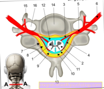
1 + 2 spinal cord - Medulla spinalis
All medical illustrations
$config[ads_text2] not foundIn contrast to the posterior strand, the long ascending fibers change to the opposite side here: they cross in the so-called commissura alba des Backmarks. That ultimately means that if that Spinal cord on the left side is damaged, the right half of the body is affected by the dysfunction.
In the case of the back-thalamic tract (Spinothalamic tract) this is the sensation of pain, temperature and rough pressure (in the sense of bruises, etc.), which are summarized as protopathic sensitivity referred to as.
Here, the lateral (lateral) of the two tracts preferentially conducts the pain and temperature stimuli and the front (anterior) rough contact and pressure stimuli.
In the elongated medulla, the medulla oblongata, the two tracts adjoin each other and run together over the thalamus, in which the third neuron is located Cerebral cortex (more precisely: to the postcentral gyrus), where conscious perception takes place.
This is where the 4th and last neuron is located.
The "pain pathway", the lateral spinothalamic tract, gets the information that it conducts to the brain from small cells in the spinal ganglion. Nociceptors, lie in the skin and mucous membranes. There are slow (C-fibers) and fast (A-δ-fibers) of the pain-conducting fibers.
$config[ads_text3] not found
A brief example may illustrate this: if we touch a hot stove top, the fast fibers report this in a flash, before we even realize it, and pull our hand away. The dull, throbbing pain that follows seconds later and lasts longer is transmitted by the slow-moving C-fibers.
There are direct contacts with im from the ascending railway Brain stem distributed core groups, which are known because of their distribution as Formatio reticularis (= ("network-like formations").
These connections lead to an increase in alertness and alertness and to stimulation of the Cardiovascular system and from Respiratory system.
The body signals to us: human, be careful - please avoid further tissue damage! Further connections in the limbic system, which is responsible for the emotional evaluation, ensure that the hot stove is remembered as negative.